Exploiting bilateral symmetry in brain lesion segmentation
Publication
Metrics
AI Quick Summary
Researchers developed a method to improve brain lesion segmentation by using bilateral symmetry in healthy brains to aid in detecting lesions. Nonlinear registration improved performance over baseline methods.
Paper Preview
Abstract
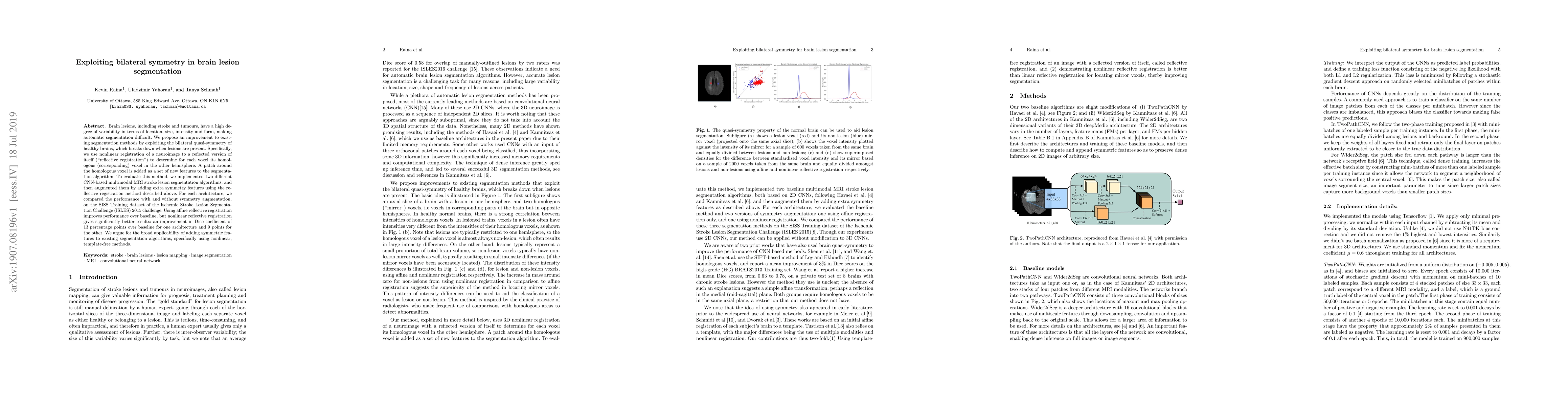
Brain lesions, including stroke and tumours, have a high degree of variability in terms of location, size, intensity and form, making automatic segmentation difficult. We propose an improvement to existing segmentation methods by exploiting the bilateral quasi-symmetry of healthy brains, which breaks down when lesions are present. Specifically, we use nonlinear registration of a neuroimage to a reflected version of itself ("reflective registration") to determine for each voxel its homologous (corresponding) voxel in the other hemisphere. A patch around the homologous voxel is added as a set of new features to the segmentation algorithm. To evaluate this method, we implemented two different CNN-based multimodal MRI stroke lesion segmentation algorithms, and then augmented them by adding extra symmetry features using the reflective registration method described above. For each architecture, we compared the performance with and without symmetry augmentation, on the SISS Training dataset of the Ischemic Stroke Lesion Segmentation Challenge (ISLES) 2015 challenge. Using affine reflective registration improves performance over baseline, but nonlinear reflective registration gives significantly better results: an improvement in Dice coefficient of 13 percentage points over baseline for one architecture and 9 points for the other. We argue for the broad applicability of adding symmetric features to existing segmentation algorithms, specifically using nonlinear, template-free methods.
AI Key Findings
Get AI-generated insights about this paper's methodology, results, significance, and more — seven facets brought into focus.
Impact
Paper Details
PDF Preview
Key Terms
Citation Network
Current paper (gray), citations (green), references (blue)
Display is limited for performance on very large graphs.
Discussion 0