Table-top extreme ultraviolet (EUV) microscopy offers unique opportunities
for label-free high resolution imaging of biological sample with
micrometer-scale penetration depth and strong elemental contrast. In this work,
we further advanced a state-of-the-art EUV ptychographic microscope and applied
it to systematic studies of the structure and composition of two prokaryotic
model bacteria, Escherichia coli and Bacillus subtilis. For the first time, a
unique combination of quantitative amplitude and phase imaging with high
precision and a record-sub-50 nm resolution has been achieved, which unveils
the ultrastructure and composition of the bacteria at a nanometer scale. This
capability allows for the clear distinction of two different bacterial types,
despite their similar size and shape. Furthermore, B. subtilis and its
endospores are examined, enabling visualization of their distinct morphology
and composition as well as detailed observations of various stages of
sporulation. We further visualized the impact of the antibiotic monazomycin on
B. subtilis for the first time, revealing its disruptive effects on cellular
structures and compositional alterations. This work demonstrates that EUV
ptychography, with a straightforward, label-free sample preparation, can serve
as a powerful, quantitative imaging modality with a unique sensitivity to
chemical information at the nanoscale, advancing biomedical studies e.g. toward
developing novel antimicrobial strategies.
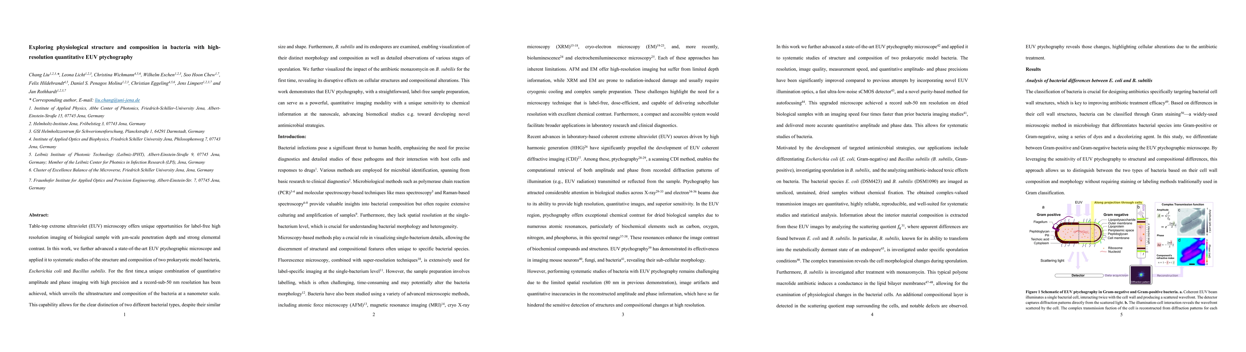
Discussion 0