This paper reviews advancements in far-field super-resolution chemical microscopy, which overcomes the diffraction limit to reveal molecular electronic or vibrational fingerprints. It highlights applications in various fields including biomedical research, material characterization, and cultural heritage conservation.
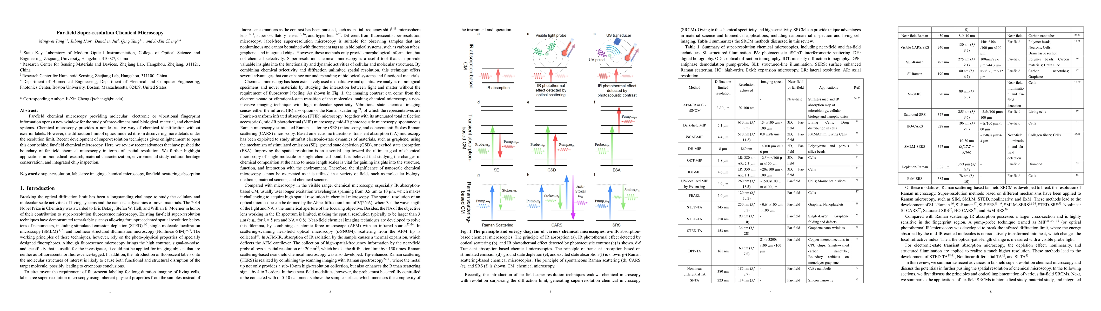
Far-field chemical microscopy providing molecular electronic or vibrational fingerprint information opens a new window for the study of three-dimensional biological, material, and chemical systems. Chemical microscopy provides a nondestructive way of chemical identification without exterior labels. However, the diffraction limit of optics hindered it from discovering more details under the resolution limit. Recent development of super-resolution techniques gives enlightenment to open this door behind far-field chemical microscopy. Here, we review recent advances that have pushed the boundary of far-field chemical microscopy in terms of spatial resolution. We further highlight applications in biomedical research, material characterization, environmental study, cultural heritage conservation, and integrated chip inspection.
Current paper (gray), citations (green), references (blue)
Tim Fingscheidt, Björn Möller, Lucas Görnhardt
Xi Lin, Manhong Yao, Junzheng Peng et al.
Jing Yu, Marc Botifoll, Jordi Arbiol et al.
Chenyu Wu, Changsheng You, Cong Zhou et al.
Title | Authors | Year | Actions |
---|---|---|---|
Applications of ultrafast nano-spectroscopy and nano-imaging with tip-based microscopy
|
Zhichen Zhao, V. Kravtsov, Zerui Wang, Zhou Zhou, Linyuan Dou, Di Huang, Zhanshan Wang, Xinbin Cheng, M. Raschke, Tao Jiang
|
2025
|
View Paper |
Advances in Super-resolution Stimulated Raman Scattering Microscopy
|
William J. Tipping, K. Faulds, D. Graham
|
2024
|
View Paper |
Surpassing the Diffraction Limit in Label-Free Optical Microscopy
|
David Palounek, M. Vala, Ł. Bujak, Ivan Kopal, Kateřina Jiříková, Yevhenii Shaidiuk, M. Piliarik
|
2024
|
View Paper |
Super-Resolution Image Reconstruction of Wavefront Coding Imaging System Based on Deep Learning Network
|
Xueyan Li, Haowen Yu, Yijian Wu, Lieshan Zhang, Di Chang, Xuhong Chu, Haoyuan Du
|
2024
|
View Paper |
Comparison of Gap-Enhanced Raman Tags and Nanoparticle Aggregates with Polarization Dependent Super-Resolution Spectral SERS Imaging.
|
Deben N. Shoup, Sanjun Fan, M. Zapata-Herrera, Hannah C. Schorr, Javier Aizpurua, Zachary D. Schultz
|
2024
|
View Paper |
SERS microscopy as a tool for comprehensive biochemical characterization in complex samples.
|
J. Kneipp, Stephan Seifert, Florian Gärber
|
2024
|
View Paper |
Far-Field Super-Resolution Microscopy Using Evanescent Illumination: A Review
|
Qianwei Zhang, Haonan Zhang, Xiaoyu Yang, Xu Liu, Mingwei Tang, Qing Yang
|
2024
|
View Paper |
Surface-Enhanced Raman Spectroscopy: Current Understanding, Challenges, and Opportunities.
|
Hao Ma, Si-Qi Pan, Wei-Li Wang, Xiaxia Yue, Xiaodong Xi, Sen Yan, Deyin Wu, Xiang Wang, Guo-kun Liu, B. Ren
|
2024
|
View Paper |
3D orientation super-resolution spatial-frequency-shift microscopy
|
Xiaowei Liu, Mingwei Tang, Ning Zhou, Chen-lei Pang, Zhong Wen, Xu Liu, Qing Yang
|
2024
|
View Paper |
Recent advances in label-free imaging and quantification techniques for the study of lipid droplets in cells.
|
Hyeonwoo Kim, Seungeun Oh, Seongsoo Lee, Kwang suk Lee, YongKeun Park
|
2023
|
View Paper |
Microsphere-assisted quantitative phase microscopy: a review
|
Vahid Abbasian, T. Pahl, Lucie Hüser, S. Lecler, Paul Montgomery, Peter Lehmann, Arash Darafsheh
|
2024
|
View Paper |
Title | Authors | Year | Actions |
---|---|---|---|
Super-resolution SRS microscopy with A-PoD
|
Hongje Jang, Yajuan Li, Anthony A. Fung, P. Bagheri, Khang Hoang, D. Skowronska-Krawczyk, Xiaoping Chen, Jane Y. Wu, B. Bintu, Lingyan Shi
|
2023
|
View Paper |
Super-resolution imaging of non-fluorescent molecules by photothermal relaxation localization microscopy
|
Pengcheng Fu, Wanlin Cao, Tianrun Chen, Xiangjie Huang, Taoran Le, Shiyao Zhu, Da-Wei Wang, Hyeon Jeong Lee, Delong Zhang
|
2023
|
View Paper |
Multicolor expansion fluorescence emission difference microscopy to reveal potential organelle contacts
|
Lu Yang, Yuran Huang, Zhimin Zhang, Yubing Han, C. Kuang
|
2022
|
View Paper |
Label-Free Autofluorescence-Detected Mid-Infrared Photothermal Microscopy of Pharmaceutical Materials.
|
Aleksandr Razumtcev, Minghe Li, Jiayue Rong, C. Teng, C. Pfluegl, L. Taylor, Garth J. Simpson
|
2022
|
View Paper |
Spatial-frequency-shift enables integrated super-resolution microscopy: advance and perspective.
|
Chen-lei Pang, Xiaowei Liu, Mingwei Tang, Qing Yang, X. Liu
|
2022
|
View Paper |
Spectral imaging with deep learning
|
Longqian Huang, Ruichen Luo, Xu Liu, X. Hao
|
2022
|
View Paper |
Bond-selective intensity diffraction tomography
|
Jian Zhao, Alex Matlock, Hongbo Zhu, Z. Song, Jiabei Zhu, Biao Wang, Fukai Chen, Yuewei Zhan, Zhicong Chen, Yihong Xu, Xingchen Lin, L. Tian, Ji-Xin Cheng
|
2022
|
View Paper |
Novel optical photothermal infrared (O-PTIR) spectroscopy for the noninvasive characterization of heritage glass-metal objects
|
A. Marchetti, V. Beltran, Gert Nuyts, Ferenc Borondics, Steven De Meyer, Marina Van Bos, J. Jaroszewicz, Elke Otten, Marjolijn Debulpaep, K. De Wael
|
2022
|
View Paper |
Micro to Nano: Multiscale IR Analyses Reveal Zinc Soap Heterogeneity in a 19th-Century Painting by Corot.
|
Xiao Ma, G. Pavlidis, E. Dillon, V. Beltran, J. Schwartz, M. Thoury, Ferenc Borondics, C. Sandt, K. Kjoller, B. Berrie, A. Centrone
|
2022
|
View Paper |
High‐Refractive‐Index Chip with Periodically Fine‐Tuning Gratings for Tunable Virtual‐Wavevector Spatial Frequency Shift Universal Super‐Resolution Imaging
|
Mingwei Tang, Yubing Han, Dehao Ye, Qianwei Zhang, Chen-lei Pang, Xiaowei Liu, Weidong Shen, Yaoguang Ma, C. Kaminski, X. Liu, Qing Yang
|
2022
|
View Paper |
Expansion microscopy opens the door to exploring more challenges
|
Mengfei Gao
|
2022
|
View Paper |
Visualizing the native cellular organization by coupling cryofixation with expansion microscopy (Cryo-ExM)
|
Marine. H. Laporte, N. Klena, V. Hamel, P. Guichard
|
2022
|
View Paper |
Super‐Resolution Vibrational Imaging Using Expansion Stimulated Raman Scattering Microscopy
|
Lixue Shi, A. Klimas, Brendan R Gallagher, Z. Cheng, Feifei Fu, P. Wijesekara, Yupeng Miao, X. Ren, Yongxin Zhao, Wei Min
|
2021
|
View Paper |
Steam disinfection releases micro(nano)plastics from silicone-rubber baby teats as examined by optical photothermal infrared microspectroscopy
|
Yu Su, Xi Hu, Hongjie Tang, Kun Lu, Huimin Li, Si-Dong Liu, B. Xing, R. Ji
|
2021
|
View Paper |
Using Infrared Photothermal Heterodyne Imaging to Characterize Micro- and Nanoplastics in Complex Environmental Matrices.
|
K. Kniazev, I. Pavlovetc, Shuang Zhang, Junyeol Kim, R. Stevenson, Kyle Doudrick, M. Kuno
|
2021
|
View Paper |
Super-resolved Raman microscopy using random structured light illumination: Concept and feasibility.
|
Hongki Lee, Hajung Yoo, Gwiyeong Moon, Kar-Ann Toh, K. Mochizuki, K. Fujita, Donghyun Kim
|
2021
|
View Paper |
Image fusion of IR and optical microscopy for mapping of biomolecules in tissue
|
Safaa Al Jedani, Conor A. Whitley, Barnaby G. Ellis, A. Triantafyllou, Caroline I Smith, P. Gunning, P. Gardner, J. Risk, P. Weightman, S. Barrett
|
2021
|
View Paper |
Correlative optical photothermal infrared and X-ray fluorescence for chemical imaging of trace elements and relevant molecular structures directly in neurons
|
Nadja Gustavsson, A. Paulus, Isak Martinsson, A. Engdahl, K. Medjoubi, K. Klementiev, A. Somogyi, T. Deierborg, Ferenc Borondics, G. Gouras, Oxana Klementieva
|
2021
|
View Paper |
Fluorescence-Detected Mid-Infrared Photothermal Microscopy.
|
Yi Zhang, Haonan Zong, Cheng Zong, Yuying Tan, Meng Zhang, Yuewei Zhan, Ji‐Xin Cheng
|
2021
|
View Paper |
Nanoscale analysis of historical paintings by means of O-PTIR spectroscopy: The identification of the organic particles in L'Arlésienne (portrait of Madame Ginoux) by Van Gogh.
|
V. Beltran, A. Marchetti, Gert Nuyts, Margje Leeuwestein, C. Sandt, Ferenc Borondics, K. De Wael
|
2021
|
View Paper |
Wide-Field Super-Resolved Raman Imaging of Carbon Materials
|
Mingqun Wang, Chonglei Zhang, S. Yan, Ting Chen, H. Fang, Xiaocong Yuan
|
2021
|
View Paper |
Quantum-enhanced nonlinear microscopy
|
Catxere A. Casacio, L. Madsen, Alex Terrasson, Muhammad Waleed, Kai Barnscheidt, B. Hage, Michael A. Taylor, W. Bowen
|
2021
|
View Paper |
Low loss and omnidirectional Si3N4 waveguide for label-free spatial frequency shift super-resolution imaging
|
Dehao Ye, Mingwei Tang, Xiaowei Liu, Yaoguang Ma, X. Liu, Qing Yang
|
2021
|
View Paper |
Direct Imaging of Integrated Circuits in CPU with 60 nm Super-Resolution Optical Microscope.
|
Guang Yang, C. Yang, Yage Chen, Boyu Yu, Yali Bi, Jiangshan Liao, Haozheng Li, Hong Wang, Yuxi Wang, Ziyu Liu, Zongsong Gan, Quan Yuan, Yi Wang, J. Xia, Ping Wang
|
2021
|
View Paper |
Background-Suppressed High-Throughput Mid-Infrared Photothermal Microscopy via Pupil Engineering.
|
Haonan Zong, Celalettin Yurdakul, Yeran Bai, Meng Zhang, M. Ünlü, Ji‐Xin Cheng
|
2021
|
View Paper |
Chip-compatible wide-field 3D nanoscopy through tunable spatial frequency shift effect
|
Xiaowei Liu, Mingwei Tang, Chao Meng, Chen-lei Pang, C. Kuang, Wei Chen, C. Kaminski, Qing Yang, X. Liu
|
2021
|
View Paper |
Scalable universal tunable virtual-wavevector spatial frequency shift (TVSFS) super-resolution imaging
|
Mingwei Tang, Yubing Han, Dehao Ye, Qianwei Zhang, Chen-lei Pang, Xiaowei Liu, Weidong Shen, Yaoguang Ma, C. Kaminski, Xu Liu, Qing Yang
|
2021
|
View Paper |
COVID-19 salivary Raman fingerprint: innovative approach for the detection of current and past SARS-CoV-2 infections
|
C. Carlomagno, D. Bertazioli, A. Gualerzi, S. Picciolini, P. Banfi, A. Lax, E. Messina, J. Navarro, L. Bianchi, A. Caronni, F. Marenco, S. Monteleone, C. Arienti, M. Bedoni
|
2021
|
View Paper |
Analysis of Fixed and Live Single Cells Using Optical Photothermal Infrared with Concomitant Raman Spectroscopy
|
A. Spadea, J. Denbigh, M. Lawrence, M. Kansiz, P. Gardner
|
2021
|
View Paper |
Vibrational Spectroscopic Detection of a Single Virus by Mid-Infrared Photothermal Microscopy.
|
Yi Zhang, Celalettin Yurdakul, A. J. Devaux, Le Wang, Xiaoji G. Xu, J. Connor, M. Ünlü, Ji‐Xin Cheng
|
2021
|
View Paper |
Ultrasensitive Vibrational Imaging of Retinoids by Visible Preresonance Stimulated Raman Scattering Microscopy
|
M. Zhuge, Kai-Chih Huang, Hyeon Jeong Lee, Ying Jiang, Yuying Tan, Haonan Lin, Pu-Ting Dong, Guangyuan Zhao, D. Matei, Qing Yang, Ji‐Xin Cheng
|
2021
|
View Paper |
Quantitative Imaging of Single Light-Absorbing Nanoparticles by Widefield Interferometric Photothermal Microscopy
|
Yu-Chien Huang, Te-Hsin Chen, Jz-Yuan Juo, S. Chu, C. Hsieh
|
2021
|
View Paper |
Imaging Isotopically Labeled Bacteria at the Single-Cell Level Using High-Resolution Optical Infrared Photothermal Spectroscopy.
|
Cassio Lima, H. Muhamadali, Yun Xu, M. Kansiz, R. Goodacre
|
2021
|
View Paper |
Super-resolution label-free volumetric vibrational imaging
|
Chenxi Qian, Kun Miao, Li-En Lin, Xinhong Chen, Jiajun Du, Lu Wei
|
2021
|
View Paper |
Discrimination of bacteria using whole organism fingerprinting: the utility of modern physicochemical techniques for bacterial typing.
|
N. AlMasoud, H. Muhamadali, Malama Chisanga, H. Alrabiah, Cassio Lima, R. Goodacre
|
2020
|
View Paper |
Surface Plasmon Localization-Based Super-resolved Raman Microscopy.
|
Hongki Lee, Kyungnam Kang, K. Mochizuki, Changhun Lee, Kar-Ann Toh, Seung Ah Lee, K. Fujita, Donghyun Kim
|
2020
|
View Paper |
Fingerprinting Bacterial Metabolic Response to Erythromycin by Raman-Integrated Mid-Infrared Photothermal Microscopy.
|
Jiabao Xu, Xiaojie Li, Zhongyue Guo, Wei E. Huang, Ji‐Xin Cheng
|
2020
|
View Paper |
Far-field transient absorption nanoscopy with sub-50 nm optical super-resolution
|
Yali Bi, C. Yang, Lei Tong, Haozheng Li, Boyu Yu, Shuai Yan, Guang Yang, Meng Deng, Yi Wang, W. Bao, Lei Ye, Ping Wang
|
2020
|
View Paper |
Far‐Field Superresolution Imaging via Spatial Frequency Modulation
|
Mingwei Tang, Xiaowei Liu, Zhong Wen, Feihong Lin, Chao Meng, X. Liu, Yaoguang Ma, Qing Yang
|
2020
|
View Paper |
Suppressing Cation Migration in Triple-Cation Lead Halide Perovskites
|
I. Pavlovetc, M. Brennan, Sergiu Draguta, Anthony Ruth, Taylor Moot, Jeffrey A. Christians, Kyle Aleshire, S. Harvey, Stefano Toso, S. Nanayakkara, Jonah F Messinger, J. Luther, M. Kuno
|
2020
|
View Paper |
High-Efficiency Perovskite Solar Cells.
|
J. Y. Kim, Jin‐Wook Lee, H. Jung, Hyunjung Shin, N. Park
|
2020
|
View Paper |
COVID-19 pathophysiology: A review
|
Koichi Yuki, M. Fujiogi, S. Koutsogiannaki
|
2020
|
View Paper |
Click-ExM enables expansion microscopy for all biomolecules
|
De-en Sun, Xinqi Fan, Yujie Shi, Hao Zhang, Zhimin Huang, Bo Cheng, Qi Tang, Wei Li, Yuntao Zhu, Jinyi Bai, Wei Liu, Yuan Li, Xiaoting Wang, Xiaoguang Lei, Xing Chen
|
2020
|
View Paper |
Evaluation of Direct Grafting Strategies via Trivalent Anchoring for Enabling Lipid Membrane and Cytoskeleton Staining in Expansion Microscopy.
|
Gang Wen, Marisa Vanheusden, Aline Acke, Donato Valli, R. Neely, Volker Leen, J. Hofkens
|
2020
|
View Paper |
Light microscopy of proteins in their ultrastructural context
|
Ons M’Saad, J. Bewersdorf
|
2020
|
View Paper |
Super‐Resolution Infrared Imaging of Polymorphic Amyloid Aggregates Directly in Neurons
|
Oxana Klementieva, C. Sandt, Isak Martinsson, M. Kansiz, G. Gouras, Ferenc Borondics
|
2020
|
View Paper |
All-digital histopathology by infrared-optical hybrid microscopy
|
M. Schnell, Shachi Mittal, Kianoush Falahkheirkhah, Anirudh Mittal, Kevin Yeh, Seth Kenkel, A. Kajdacsy-Balla, P. Carney, R. Bhargava
|
2020
|
View Paper |
Higher-order coherent anti-Stokes Raman scattering microscopy realizes label-free super-resolution vibrational imaging
|
Li Gong, Wei Zheng, Ying Ma, Zhiwei Huang
|
2020
|
View Paper |
Super-resolution fluorescence-assisted diffraction computational tomography reveals the three-dimensional landscape of the cellular organelle interactome
|
Dashan Dong, Xiaoshuai Huang, Liuju Li, Heng Mao, Yanquan Mo, Guangyi Zhang, Zhe Zhang, Jiayu Shen, Wei Liu, Zeming Wu, Guanghui Liu, Yanmei Liu, Hong Yang, Q. Gong, K. Shi, Liangyi Chen
|
2020
|
View Paper |
Subcellular Chemical Imaging: New Avenues in Cell Biology.
|
J. Decelle, G. Veronesi, B. Gallet, H. Stryhanyuk, Pietro Benettoni, Matthias Schmidt, R. Tucoulou, M. Passarelli, S. Bohic, P. Clode, N. Musat
|
2020
|
View Paper |
Label-free metabolic imaging by mid-infrared optoacoustic microscopy in living cells
|
M. Pleitez, Asrar Ali Khan, A. Soldá, Andriy Chmyrov, Josefine Reber, Francesca Gasparin, M. Seeger, Benedikt Schätz, S. Herzig, M. Scheideler, V. Ntziachristos
|
2019
|
View Paper |
Label-free biochemical quantitative phase imaging with mid-infrared photothermal effect
|
Miu Tamamitsu, Keiichiro Toda, Hiroyuki Shimada, Takaaki Honda, Masaharu Takarada, K. Okabe, Yu Nagashima, R. Horisaki, T. Ideguchi
|
2019
|
View Paper |
High-speed in vitro intensity diffraction tomography
|
Jiaji Li, Alex Matlock, Yunzhe Li, Qian Chen, Chao Zuo, L. Tian
|
2019
|
View Paper |
Expansion Microscopy of Lipid Membranes
|
Emmanouil D. Karagiannis, Jeong Seuk Kang, T. Shin, Amauche Emenari, Shoh M. Asano, Leanne Lin, Emma K. Costa, A. Marblestone, N. Kasthuri, E. Boyden
|
2019
|
View Paper |
Three-dimensional imaging of integrated circuits with macro- to nanoscale zoom
|
M. Holler, M. Odstrčil, M. Guizar‐Sicairos, M. Lebugle, Elisabeth Müller, S. Finizio, Gemma Tinti, C. David, Joshua Zusman, W. Unglaub, O. Bunk, J. Raabe, A. Levi, G. Aeppli
|
2019
|
View Paper |
High-resolution 3D refractive index microscopy of multiple-scattering samples from intensity images.
|
Shwetadwip Chowdhury, Michael Chen, Regina Eckert, D. Ren, Fan Wu, N. Repina, L. Waller
|
2019
|
View Paper |
Biological imaging of chemical bonds by stimulated Raman scattering microscopy
|
Fanghao Hu, Lixue Shi, Wei Min
|
2019
|
View Paper |
Molecular organization of mammalian meiotic chromosome axis revealed by expansion STORM microscopy
|
Huizhong Xu, Zhisong Tong, Qing Ye, Tengqian Sun, Zhenmin Hong, Lunfeng Zhang, A. Bortnick, Sunglim Cho, P. Beuzer, Josh Axelrod, Qiongzheng Hu, Melissa Wang, S. Evans, C. Murre, Li-Fan Lu, Shan Sun, K. Corbett, Hu Cang
|
2019
|
View Paper |
Label-Free Optical Nanoscopy of Single Layer Graphene.
|
Giulia Zanini, K. Korobchevskaya, T. Deguchi, A. Diaspro, P. Bianchini
|
2019
|
View Paper |
Fingerprinting a Living Cell by Raman Integrated Mid-Infrared Photothermal Microscopy.
|
Xiaojie Li, Delong Zhang, Yeran Bai, Weibiao Wang, Jingqiu Liang, Ji‐Xin Cheng
|
2019
|
View Paper |
Imaging with quantum states of light
|
P. Moreau, E. Toninelli, T. Gregory, M. Padgett
|
2019
|
View Paper |
Amplified Expansion Stimulated Emission Depletion Microscopy
|
Doyeon Kim, Taeyeong Kim, Jooyong Lee, S. Shim
|
2019
|
View Paper |
High-resolution, high-contrast mid-infrared imaging of fresh biological samples with ultraviolet-localized photoacoustic microscopy
|
Junhui Shi, T. T. Wong, Yun He, Lei Li, Ruiying Zhang, C. Yung, Jeeseong Hwang, K. Maslov, Lihong V. Wang
|
2019
|
View Paper |
On‐Chip Super‐Resolution Imaging with Fluorescent Polymer Films
|
Chen-lei Pang, Jingxi Li, Mingwei Tang, Jianpu Wang, I. Mela, Florian Ströhl, Lisa Hecker, Weidong Shen, Qiulan Liu, Xiaowei Liu, Yinan Wang, Hao Zhang, Mengnan Xu, Xinghong Zhang, X. Liu, Qing Yang, C. Kaminski
|
2019
|
View Paper |
Saturated Stimulated-Raman-Scattering Microscopy for Far-Field Superresolution Vibrational Imaging
|
Li Gong, Wei Zheng, Ying Ma, Zhiwei Huang
|
2019
|
View Paper |
Quantitative phase imaging with molecular vibrational sensitivity.
|
Miu Tamamitsu, Keiichiro Toda, R. Horisaki, T. Ideguchi
|
2019
|
View Paper |
Structured illumination microscopy using a photonic chip
|
Ø. Helle, F. Dullo, Marcel Lahrberg, Jean-Claude Tinguely, O. Hellesø, B. Ahluwalia
|
2019
|
View Paper |
Selective suppression of CARS signal with two competing stimulated Raman scattering processes.
|
B. J. Rao, D. Choi, M. Cho
|
2018
|
View Paper |
Expansion microscopy: principles and uses in biological research
|
A. Wassie, Yongxin Zhao, E. Boyden
|
2018
|
View Paper |
Imaging cellular ultrastructures using expansion microscopy (U-ExM)
|
D. Gambarotto, Fabian Zwettler, M. Le Guennec, Marketa Schmidt-Cernohorska, Denis Fortun, S. Borgers, Jörn Heine, Jan-Gero Schloetel, M. Reuss, M. Unser, E. Boyden, M. Sauer, V. Hamel, P. Guichard
|
2018
|
View Paper |
Ultrafast chemical imaging by widefield photothermal sensing of infrared absorption
|
Yeran Bai, Delong Zhang, L. Lan, Yimin Huang, K. Maize, A. Shakouri, Ji‐Xin Cheng
|
2018
|
View Paper |
Applications of nanostructures in wide-field, label-free super resolution microscopy
|
Xiaowei Liu, Chao Meng, Xuechu Xu, Mingwei Tang, Chen-lei Pang, Qing Yang
|
2018
|
View Paper |
Near-resonance enhanced label-free stimulated Raman scattering microscopy with spatial resolution near 130 nm
|
Yali Bi, C. Yang, Yage Chen, Shuai Yan, Guang Yang, Yaozu Wu, Guoping Zhang, Ping Wang
|
2018
|
View Paper |
Real-time high-resolution mid-infrared optical coherence tomography
|
N. Israelsen, C. Petersen, A. Barh, D. Jain, Mikkel Jensen, G. Hannesschläger, P. Tidemand‐Lichtenberg, C. Pedersen, A. Podoleanu, O. Bang
|
2018
|
View Paper |
Quantitative phase imaging in biomedicine
|
Yongkeun Park, C. Depeursinge, G. Popescu
|
2018
|
View Paper |
Combined expansion microscopy with structured illumination microscopy for analyzing protein complexes
|
Yongfu Wang, Zulin Yu, Cori K. Cahoon, T. Parmely, Nancy Thomas, J. Unruh, Brian D. Slaughter, R. Hawley
|
2018
|
View Paper |
Selective suppression of CARS signal with three-beam competing stimulated Raman scattering processes.
|
D. Choi, B. J. Rao, Doyeon Kim, S. Shim, Hanju Rhee, M. Cho
|
2018
|
View Paper |
NanoSIMS for biological applications: Current practices and analyses.
|
Jamie R. Nuñez, R. Renslow, J. Cliff, C. Anderton
|
2018
|
View Paper |
Expansion stimulated emission depletion microscopy (ExSTED)
|
Mengfei Gao, Riccardo Maraspini, O. Beutel, Amin Zehtabian, B. Eickholt, A. Honigmann, H. Ewers
|
2018
|
View Paper |
High-Speed Spectroscopic Transient Absorption Imaging of Defects in Graphene.
|
Kai-Chih Huang, J. McCall, Pu Wang, Chien-Sheng Liao, Gregory S. Eakins, Ji‐Xin Cheng, Chen Yang
|
2018
|
View Paper |
Subdiffraction Infrared Imaging of Mixed Cation Perovskites: Probing Local Cation Heterogeneities
|
R. Chatterjee, I. Pavlovetc, Kyle Aleshire, G. Hartland, M. Kuno
|
2018
|
View Paper |
STED super-resolved microscopy
|
G. Vicidomini, P. Bianchini, A. Diaspro
|
2018
|
View Paper |
High-speed optical coherence tomography by circular interferometric ranging
|
M. Siddiqui, A. Nam, S. Tozburun, N. Lippok, C. Blatter, B. Vakoc
|
2017
|
View Paper |
Realization of Wafer-Scale Hyperlens Device for Sub-diffractional Biomolecular Imaging
|
Dasol Lee, Yangdoo Kim, Minkyung Kim, Sunae So, H. Choi, J. Mun, D. Nguyen, Trevon Badloe, J. Ok, Kyunghoon Kim, Heon Lee, J. Rho
|
2017
|
View Paper |
Three-dimensional wide-field pump-probe structured illumination microscopy.
|
Yang-Hyo Kim, P. So
|
2017
|
View Paper |
Bond-Selective Imaging of Cells by Mid-Infrared Photothermal Microscopy in High Wavenumber Region.
|
Yeran Bai, Delong Zhang, Chen Li, Cheng Liu, Ji‐Xin Cheng
|
2017
|
View Paper |
Super-Resolution Far-Field Infrared Imaging by Photothermal Heterodyne Imaging.
|
Zhongming Li, Kyle Aleshire, M. Kuno, G. Hartland
|
2017
|
View Paper |
Nanoscale imaging of clinical specimens using pathology-optimized expansion microscopy
|
Yongxin Zhao, Octavian Bucur, H. Irshad, Fei Chen, Astrid Weins, A. Stancu, Eun-Young Oh, Marcello DiStasio, Vanda F Torous, B. Glass, I. Stillman, S. Schnitt, Andrew H. Beck, E. Boyden
|
2017
|
View Paper |
Spatial-domain filter enhanced subtraction microscopy and application to mid-IR imaging.
|
Mahendar Kumbham, R. Mouras, A. Mani, S. Daly, K. O’Dwyer, A. Toma, P. Bianchini, A. Diaspro, Ning Liu, S. Tofail, C. Silien
|
2017
|
View Paper |
Mid-Infrared Photothermal Imaging of Active Pharmaceutical Ingredients at Submicrometer Spatial Resolution.
|
Chen Li, Delong Zhang, M. Slipchenko, Ji‐Xin Cheng
|
2017
|
View Paper |
Stimulated Raman Scattering: From Bulk to Nano.
|
Richard C. Prince, R. Frontiera, E. Potma
|
2017
|
View Paper |
Iterative expansion microscopy
|
Jae‐Byum Chang, Fei Chen, Young-Gyu Yoon, Erica E. Jung, H. Babcock, Jeong Seuk Kang, Shoh M. Asano, Ho‐Jun Suk, N. Pak, P. Tillberg, A. Wassie, Dawen Cai, E. Boyden
|
2017
|
View Paper |
High-resolution non-destructive three-dimensional imaging of integrated circuits
|
M. Holler, M. Guizar‐Sicairos, E. Tsai, R. Dinapoli, Elisabeth Müller, O. Bunk, J. Raabe, G. Aeppli
|
2017
|
View Paper |
Stimulated Emission Depletion Microscopy.
|
H. Blom, J. Widengren
|
2017
|
View Paper |
Fluorescent Nanowire Ring Illumination for Wide-Field Far-Field Subdiffraction Imaging.
|
Xiaowei Liu, C. Kuang, X. Hao, Chen-lei Pang, Pengfei Xu, Haifeng Li, Y. Liu, Chaojian Yu, Yingke Xu, Di Nan, Weidong Shen, Yue Fang, Lenian He, X. Liu, Qing Yang
|
2017
|
View Paper |
Refractive index tomography with structured illumination
|
Shwetadwip Chowdhury, W. Eldridge, A. Wax, J. Izatt
|
2017
|
View Paper |
A Supercritical Lens Optical Label‐Free Microscopy: Sub‐Diffraction Resolution and Ultra‐Long Working Distance
|
Fei Qin, Kun Huang, Jianfeng Wu, J. Teng, C. Qiu, M. Hong
|
2017
|
View Paper |
Scanning superlens microscopy for non-invasive large field-of-view visible light nanoscale imaging
|
Feifei Wang, Lianqing Liu, Haibo Yu, Yangdong Wen, Peng Yu, Zhu Liu, Yuechao Wang, W. Li
|
2016
|
View Paper |
Doubling the far-field resolution in mid-infrared microscopy.
|
Mahendar Kumbham, S. Daly, K. O’Dwyer, R. Mouras, Ning Liu, A. Mani, A. Peremans, S. Tofail, C. Silien
|
2016
|
View Paper |
Imaging theory of structured pump-probe microscopy.
|
Eric S. Massaro, Andrew H. Hill, Casey L. Kennedy, Erik M. Grumstrup
|
2016
|
View Paper |
Depth-resolved mid-infrared photothermal imaging of living cells and organisms with submicrometer spatial resolution
|
Delong Zhang, Chen Li, Chi Zhang, M. Slipchenko, Gregory S. Eakins, Ji‐Xin Cheng
|
2016
|
View Paper |
Copper Uptake, Intracellular Localization, and Speciation in Marine Microalgae Measured by Synchrotron Radiation X-ray Fluorescence and Absorption Microspectroscopy.
|
M. Adams, C. T. Dillon, S. Vogt, B. Lai, J. Stauber, D. Jolley
|
2016
|
View Paper |
Multiplexed and scalable super-resolution imaging of three-dimensional protein localization in size-adjustable tissues
|
T. Ku, Justin Swaney, Jeong-Yoon Park, A. Albanese, Evan Murray, J. H. Cho, Young-Gyun Park, Vamsi Mangena, Jiapei Chen, Kwanghun Chung
|
2016
|
View Paper |
Protein-retention expansion microscopy of cells and tissues labeled using standard fluorescent proteins and antibodies
|
P. Tillberg, Fei Chen, Kiryl D Piatkevich, Yongxin Zhao, C. Yu, Brian P. English, L. Gao, Anthony J. Martorell, Ho‐Jun Suk, Fumiaki Yoshida, Ellen M. DeGennaro, Douglas H. Roossien, Guanyu Gong, Uthpala I. Seneviratne, S. Tannenbaum, R. Desimone, Dawen Cai, E. Boyden
|
2016
|
View Paper |
Nanoscale Imaging of RNA with Expansion Microscopy
|
Fei Chen, A. Wassie, Allison Coté, Anubhav Sinha, Shahar Alon, Shoh M. Asano, Evan R. Daugharthy, Jae‐Byum Chang, A. Marblestone, G. Church, A. Raj, E. Boyden
|
2016
|
View Paper |
Expansion microscopy with conventional antibodies and fluorescent proteins
|
Tyler J. Chozinski, Aaron R. Halpern, Haruhisa Okawa, Hyeon-Jin Kim, G. J. Tremel, R. Wong, J. Vaughan
|
2016
|
View Paper |
Super-Resolution Structured Pump–Probe Microscopy
|
Eric S. Massaro, Andrew H. Hill, Erik M. Grumstrup
|
2016
|
View Paper |
Super-Resolution Chemical Imaging with Plasmonic Substrates
|
Aeli P Olson, C. T. Ertsgaard, Sarah N. Elliott, N. Lindquist
|
2016
|
View Paper |
Far-Field Subdiffraction Imaging of Semiconductors Using Nonlinear Transient Absorption Differential Microscopy
|
Ning Liu, Mahendar Kumbham, Isabel A. Pita, Yina Guo, P. Bianchini, A. Diaspro, S. Tofail, A. Peremans, C. Silien
|
2016
|
View Paper |
Toward Label-Free Super-Resolution Microscopy
|
Ruchira Silva, Christian T. Graefe, R. Frontiera
|
2016
|
View Paper |
Structured line illumination Raman microscopy
|
Kozue Watanabe, A. Palonpon, N. Smith, Liang-da Chiu, A. Kasai, Hitoshi Hashimoto, S. Kawata, K. Fujita
|
2015
|
View Paper |
Vibrational spectroscopic imaging of living systems: An emerging platform for biology and medicine
|
Ji‐Xin Cheng, X. Xie
|
2015
|
View Paper |
Diffraction tomography with Fourier ptychography.
|
R. Horstmeyer, J. Chung, X. Ou, G. Zheng, Changhuei Yang
|
2015
|
View Paper |
New modes in label-free super resolution based on photo-modulated reflectivity.
|
O. Tzang, O. Cheshnovsky
|
2015
|
View Paper |
Infrared nanospectroscopy characterization of oligomeric and fibrillar aggregates during amyloid formation
|
F. Ruggeri, G. Longo, S. Faggiano, E. Lipiec, A. Pastore, G. Dietler
|
2015
|
View Paper |
Super-Spatial- and -Spectral-Resolution in Vibrational Imaging via Saturated Coherent Anti-Stokes Raman Scattering
|
Y. Yonemaru, A. Palonpon, S. Kawano, N. Smith, S. Kawata, K. Fujita
|
2015
|
View Paper |
Expansion microscopy
|
Fei Chen, P. Tillberg, E. Boyden
|
2015
|
View Paper |
Super-resolution in label-free photomodulated reflectivity.
|
O. Tzang, A. Pevzner, R. Marvel, R. Haglund, O. Cheshnovsky
|
2015
|
View Paper |
Dynamic placement of plasmonic hotspots for super-resolution surface-enhanced Raman scattering.
|
C. T. Ertsgaard, Rachel M. McKoskey, Isabel S Rich, N. Lindquist
|
2014
|
View Paper |
Elemental and Chemically Specific X-ray Fluorescence Imaging of Biological Systems
|
M. J. Pushie, I. Pickering, M. Korbas, M. Hackett, Graham N George
|
2014
|
View Paper |
Breaking the diffraction limit by saturation in stimulated-Raman-scattering microscopy: A theoretical study
|
Li Gong, Haifeng Wang
|
2014
|
View Paper |
A method for super-resolved CARS microscopy with structured illumination in two dimensions.
|
J. Park, Sang-Won Lee, Eun Seong Lee, J. Y. Lee
|
2014
|
View Paper |
Enhancing spatial resolution in digital holographic microscopy by biprism structured illumination.
|
E. Sánchez-Ortiga, M. Martínez-Corral, G. Saavedra, J. Garcia-Sucerquia
|
2014
|
View Paper |
Live-cell imaging of alkyne-tagged small biomolecules by stimulated Raman scattering
|
Lu Wei, Fanghao Hu, Yihui Shen, Zhixing Chen, Yong Yu, C. Lin, Meng C. Wang, Wei Min
|
2014
|
View Paper |
A 1.7 nm resolution chemical analysis of carbon nanotubes by tip-enhanced Raman imaging in the ambient
|
Chi Chen, N. Hayazawa, S. Kawata
|
2014
|
View Paper |
Super-resolution nonlinear photothermal microscopy.
|
D. Nedosekin, E. Galanzha, E. Dervishi, A. Biris, V. Zharov
|
2014
|
View Paper |
Label-Free Nanometer-Resolution Imaging of Biological Architectures through Surface Enhanced Raman Scattering
|
Sencer Ayas, G. Cinar, Alper D Ozkan, Z. Soran, O. Ekiz, D. Kocaay, A. Tomak, P. Toren, Y. Kaya, Ilknur Tunc, H. Zareie, T. Tekinay, A. Tekinay, M. O. Guler, A. Dana
|
2013
|
View Paper |
Far-field Imaging of Non-fluorescent Species with Sub-diffraction Resolution
|
Pu Wang, M. Slipchenko, James R Mitchell, Chen Yang, E. Potma, Xianfan Xu, Ji‐Xin Cheng
|
2013
|
View Paper |
Chemical imaging beyond the diffraction limit: experimental validation of the PTIR technique.
|
B. Lahiri, G. Holland, A. Centrone
|
2013
|
View Paper |
AFM–IR: Combining Atomic Force Microscopy and Infrared Spectroscopy for Nanoscale Chemical Characterization
|
A. Dazzi, C. Prater, Q. Hu, D. Chase, J. Rabolt, C. Marcott
|
2012
|
View Paper |
Structured oblique illumination microscopy for enhanced resolution imaging of non-fluorescent, coherently scattering samples
|
Shwetadwip Chowdhury, Al-Hafeez Z. Dhalla, J. Izatt
|
2012
|
View Paper |
A super-oscillatory lens optical microscope for subwavelength imaging.
|
E. Rogers, J. Lindberg, T. Roy, S. Savo, J. Chad, Mark R. Dennis, N. Zheludev
|
2012
|
View Paper |
Hyperlenses and metalenses for far-field super-resolution imaging
|
Dylan Lu, Zhaowei Liu
|
2012
|
View Paper |
Nonlinear structured-illumination microscopy with a photoswitchable protein reveals cellular structures at 50-nm resolution
|
E. Rego, L. Shao, J. J. Macklin, Lukman Winoto, G. Johansson, Nicholas Kamps-Hughes, M. Davidson, M. Gustafsson
|
2011
|
View Paper |
Spherical hyperlens for two-dimensional sub-diffractional imaging at visible frequencies.
|
J. Rho, Ziliang Ye, Y. Xiong, Xiaobo Yin, Zhaowei Liu, Hyeunseok Choi, G. Bartal, Xiang Zhang
|
2010
|
View Paper |
Nanoscale chemical imaging using top-illumination tip-enhanced Raman spectroscopy.
|
Johannes Stadler, T. Schmid, Renato Zenobi
|
2010
|
View Paper |
Room-Temperature Detection of a Single Molecule’s Absorption by Photothermal Contrast
|
A. Gaiduk, M. Yorulmaz, Paul V. Ruijgrok, M. Orrit
|
2010
|
View Paper |
Optical virtual imaging at 50 nm lateral resolution with a white-light nanoscope.
|
Zengbo Wang, Wei Guo, Lin Li, B. Luk’yanchuk, Ashfaq Khan, Zhu Liu, Zaichun Chen, M. Hong
|
2010
|
View Paper |
Principles and techniques of digital holographic microscopy
|
Myung K. Kim
|
2010
|
View Paper |
Super-resolution imaging with small organic fluorophores.
|
M. Heilemann, S. van de Linde, Anindita Mukherjee, M. Sauer
|
2009
|
View Paper |
Near-field focusing and magnification through self-assembled nanoscale spherical lenses
|
J. Y. Lee, B. Hong, W. Kim, S. K. Min, Yukyung Kim, M. Jouravlev, R. Bose, K. Kim, I. Hwang, L. Kaufman, C. Wong, P. Kim, Kwang S. Kim
|
2009
|
View Paper |
High-speed atomic force microscopy for nano-visualization of dynamic biomolecular processes
|
T. Ando, T. Uchihashi, T. Fukuma
|
2008
|
View Paper |
From Bulk to Nano
|
Carmen-Gabriela Stefanita
|
2008
|
View Paper |
Subdiffraction-resolution fluorescence imaging with conventional fluorescent probes.
|
M. Heilemann, S. van de Linde, M. Schüttpelz, R. Kasper, B. Seefeldt, Anindita Mukherjee, Philip Tinnefeld, M. Sauer
|
2008
|
View Paper |
Tip-enhanced Raman spectroscopy and microscopy on single dye molecules with 15 nm resolution.
|
Jens Steidtner, B. Pettinger
|
2008
|
View Paper |
Three-dimensional sub–100 nm resolution fluorescence microscopy of thick samples
|
M. Juette, T. Gould, M. Lessard, Michael J. Mlodzianoski, B. S. Nagpure, B. Bennett, S. Hess, J. Bewersdorf
|
2008
|
View Paper |
Development of optical hyperlens for imaging below the diffraction limit.
|
Hyesog Lee, Zhaowei Liu, Y. Xiong, Cheng Sun, Xiang Zhang
|
2007
|
View Paper |
Ultra-high resolution imaging by fluorescence photoactivation localization microscopy.
|
S. Hess, T. P. K. Girirajan, Michael D. Mason
|
2006
|
View Paper |
Sub-diffraction-limit imaging by stochastic optical reconstruction microscopy (STORM)
|
Michael J Rust, Mark Bates, X. Zhuang
|
2006
|
View Paper |
Imaging Intracellular Fluorescent Proteins at Nanometer Resolution
|
E. Betzig, G. Patterson, R. Sougrat, O. W. Lindwasser, S. Olenych, J. Bonifacino, M. Davidson, J. Lippincott-Schwartz, H. Hess
|
2006
|
View Paper |
Macromolecular-scale resolution in biological fluorescence microscopy.
|
Gerald Donnert, J. Keller, R. Medda, M. Andrei, S. Rizzoli, R. Lührmann, R. Jahn, C. Eggeling, S. Hell
|
2006
|
View Paper |
STED microscopy reveals that synaptotagmin remains clustered after synaptic vesicle exocytosis
|
K. Willig, S. Rizzoli, V. Westphal, R. Jahn, S. Hell
|
2006
|
View Paper |
Cell refractive index tomography by digital holographic microscopy.
|
F. Charriére, A. Marian, F. Montfort, Jonas Kuehn, T. Colomb, E. Cuche, P. Marquet, C. Depeursinge
|
2006
|
View Paper |
Force measurements with the atomic force microscope: Technique, interpretation and applications
|
H. Butt, B. Cappella, M. Kappl
|
2005
|
View Paper |
Local infrared microspectroscopy with subwavelength spatial resolution with an atomic force microscope tip used as a photothermal sensor.
|
A. Dazzi, R. Prazeres, F. Glotin, J. Ortega
|
2005
|
View Paper |
Nonlinear structured-illumination microscopy: wide-field fluorescence imaging with theoretically unlimited resolution.
|
M. Gustafsson
|
2005
|
View Paper |
Photothermal heterodyne imaging of individual nonfluorescent nanoclusters and nanocrystals.
|
S. Berciaud, L. Cognet, G. A. Blab, B. Lounis
|
2004
|
View Paper |
High-resolution near-field Raman microscopy of single-walled carbon nanotubes.
|
A. Hartschuh, E. Sánchez, X. S. Xie, L. Novotný
|
2003
|
View Paper |
Photothermal Imaging of Nanometer-Sized Metal Particles Among Scatterers
|
D. Boyer, P. Tamarat, A. Maali, B. Lounis, M. Orrit
|
2002
|
View Paper |
Electron Microscopy and Analysis
|
K. Knowles
|
2001
|
View Paper |
Vibrational Imaging with High Sensitivity via Epidetected Coherent Anti-Stokes Raman Scattering Microscopy
|
A. Volkmer, Ji‐Xin Cheng, X. Xie
|
2001
|
View Paper |
Surpassing the lateral resolution limit by a factor of two using structured illumination microscopy
|
M. Gustafsson
|
2000
|
View Paper |
Near-field probing of vibrational absorption for chemical microscopy
|
B. Knoll, F. Keilmann
|
1999
|
View Paper |
Laterally modulated excitation microscopy: improvement of resolution by using a diffraction grating
|
R. Heintzmann, C. Cremer
|
1999
|
View Paper |
Fourier transform spectroscopic imaging using an infrared focal-plane array detector.
|
Lewis En, P. Treado, Reeder Rc, G. M. Story, A. E. Dowrey, C. Marcott, I. W. Levin
|
1995
|
View Paper |
Breaking the diffraction resolution limit by stimulated emission: stimulated-emission-depletion fluorescence microscopy.
|
S. Hell, Jan Wichmann
|
1994
|
View Paper |
Thermal lens technique: a new method of absorption spectroscopy.
|
M. E. Long, R. Swofford, A. C. Albrecht
|
1976
|
View Paper |
Photo induced Force Microscopy: chemical spectroscopy beyond the diffraction limit
|
Philip R. Davies, Josh Davies-Jones
|
2022
|
View Paper |
: A Review of the
|
Michael Smith, David Brink
|
2018
|
View Paper |
High-resolution three-dimensional tomographic diffractive microscopy of transparent inorganic and biological samples.
|
M. Debailleul, V. Georges, B. Simon, R. Morin, O. Haeberlé
|
2009
|
View Paper |
Current Applications of Liquid Chromatography / Mass Spectrometry in Pharmaceutical Discovery After a Decade of Innovation
|
Jeremy K. Nicholson, M. Mirkin, Jane A. Dickerson, Colin D. Whitmore, Emily H. Turner, M. Palcic, Ole Hindsgaul, N. Dovichi, N. L. Allbritton
|
2008
|
View Paper |
Femtosecond stimulated Raman spectroscopy.
|
P. Kukura, D. McCamant, R. Mathies
|
2007
|
View Paper |
Micro to Nano: Multiscale IR Analyses Reveal Zinc Soap Heterogeneity in a 19th-Century Painting by Corot
|
View Paper | ||
Theoretical study
|
Dr. Kadhim A. Hubeatir
|
View Paper |
Showing first page preview. Click "Open Full PDF" to view the complete paper.
Your comment contains inappropriate language and has been rejected.
Are you sure you want to remove this tag? This action cannot be undone.
Someone just favorited a paper!
Comments (0)