Academic Profile
Statistics
Similar Authors
Papers on arXiv
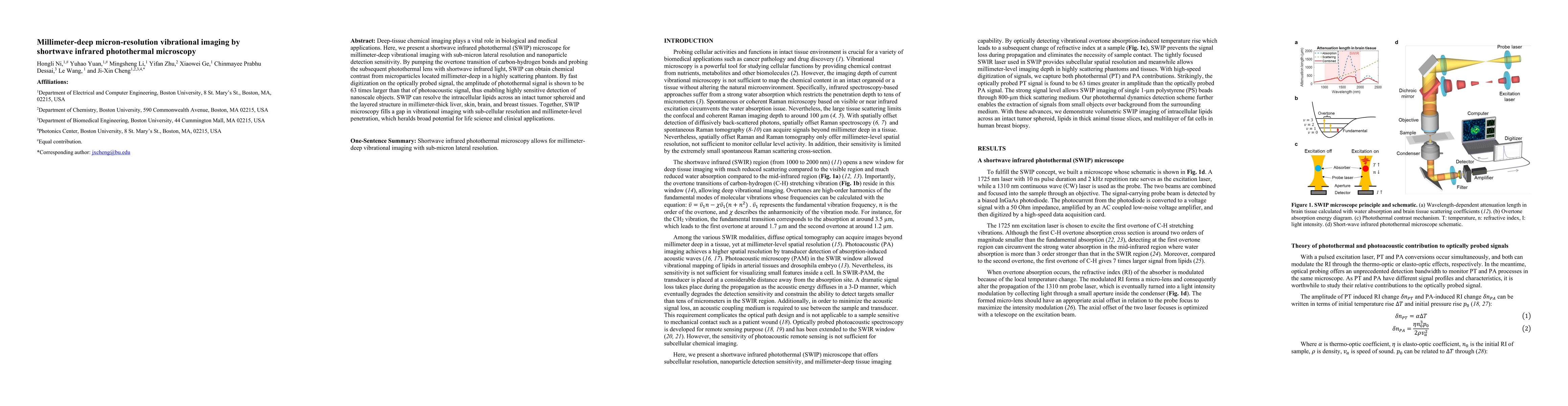
Deep-tissue chemical imaging plays a vital role in biological and medical applications. Here, we present a shortwave infrared photothermal (SWIP) microscope for millimeter-deep vibrational imaging w...
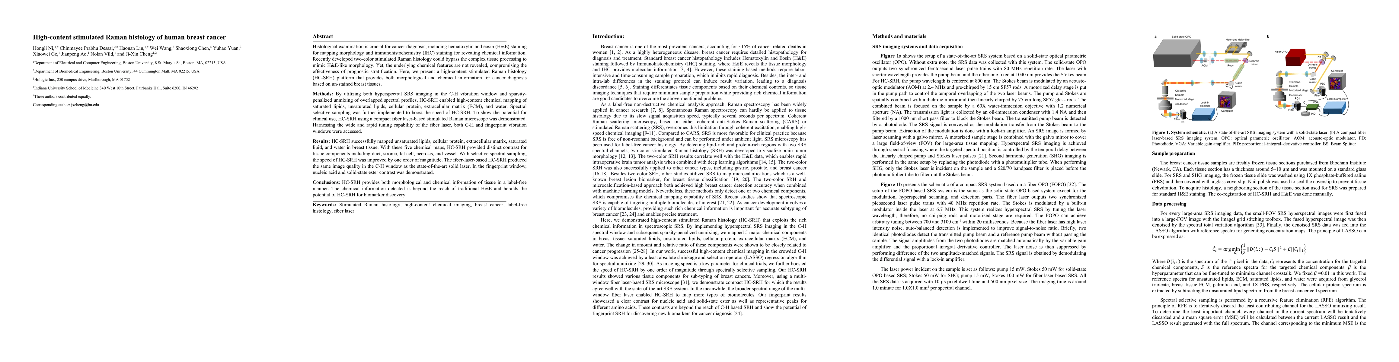
Histological examination is crucial for cancer diagnosis, including hematoxylin and eosin (H&E) staining for mapping morphology and immunohistochemistry (IHC) staining for revealing chemical informa...
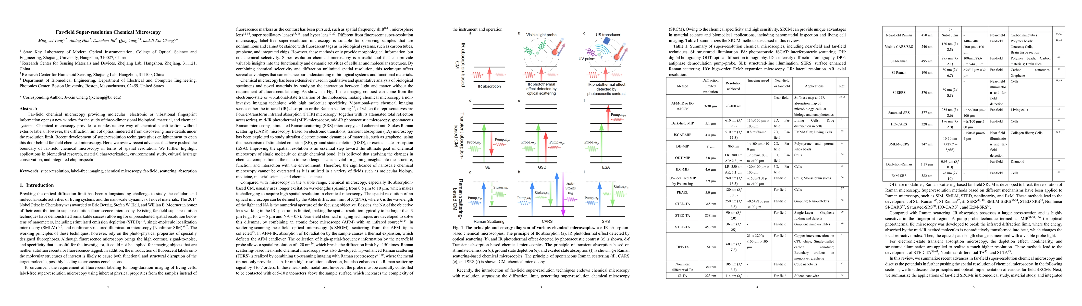
Far-field chemical microscopy providing molecular electronic or vibrational fingerprint information opens a new window for the study of three-dimensional biological, material, and chemical systems. ...
Amyloid proteins are associated with a broad spectrum of neurodegenerative diseases. However, it remains a grand challenge to extract molecular structure information from intracellular amyloid prote...
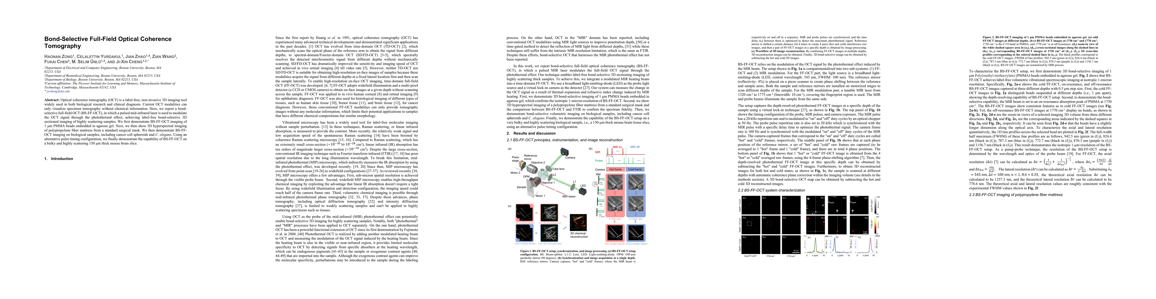
Optical coherence tomography (OCT) is a label-free, non-invasive 3D imaging tool widely used in both biological research and clinical diagnosis. Current OCT modalities can only visualize specimen to...
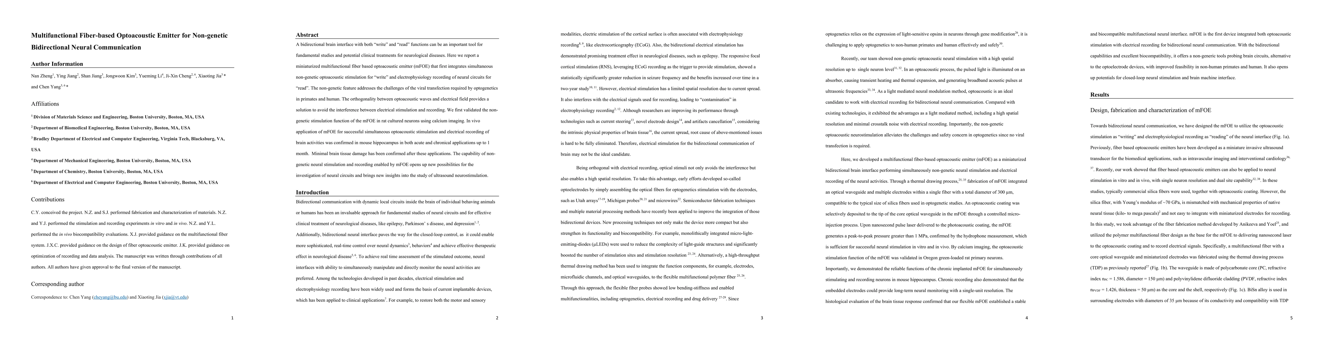
A bidirectional brain interface with both "write" and "read" functions can be an important tool for fundamental studies and potential clinical treatments for neurological diseases. Here we report a ...
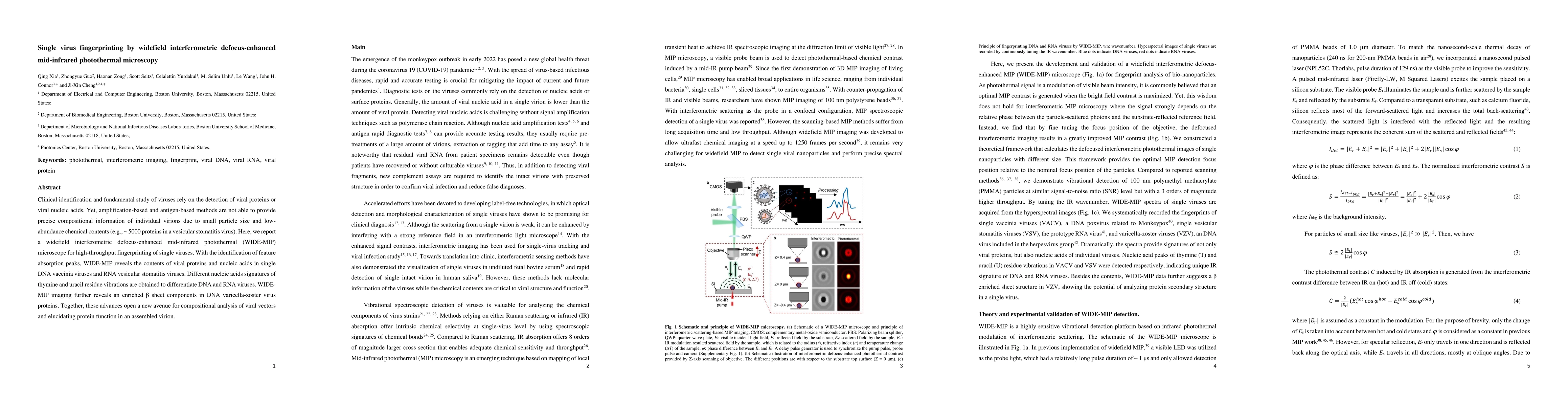
Clinical identification and fundamental study of viruses rely on the detection of viral proteins or viral nucleic acids. Yet, amplification-based and antigen-based methods are not able to provide pr...
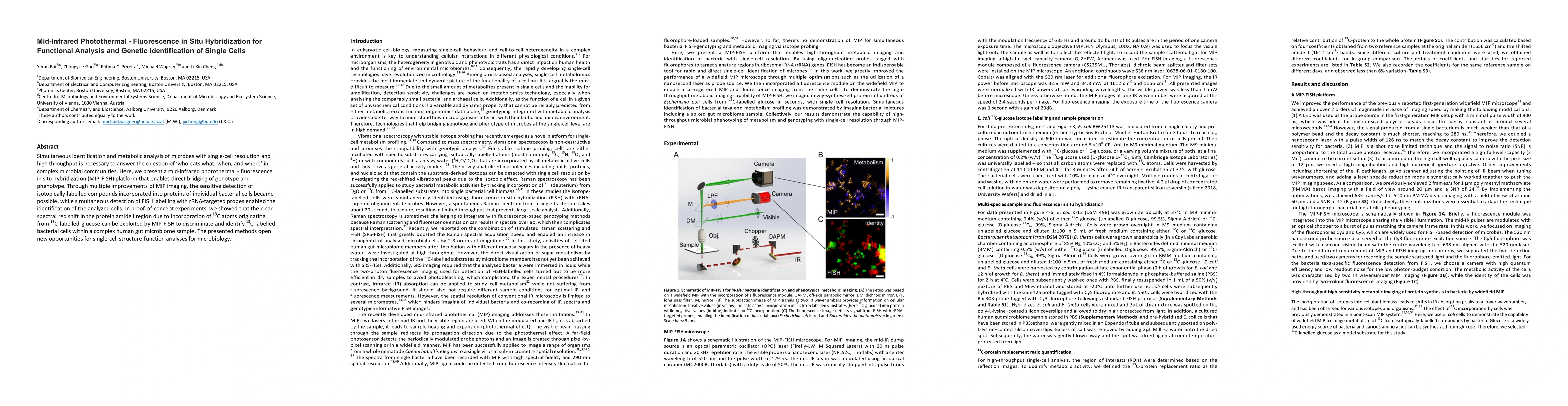
Simultaneous identification and metabolic analysis of microbes with single-cell resolution and high throughput is necessary to answer the question of "who eats what, when, and where" in complex micr...
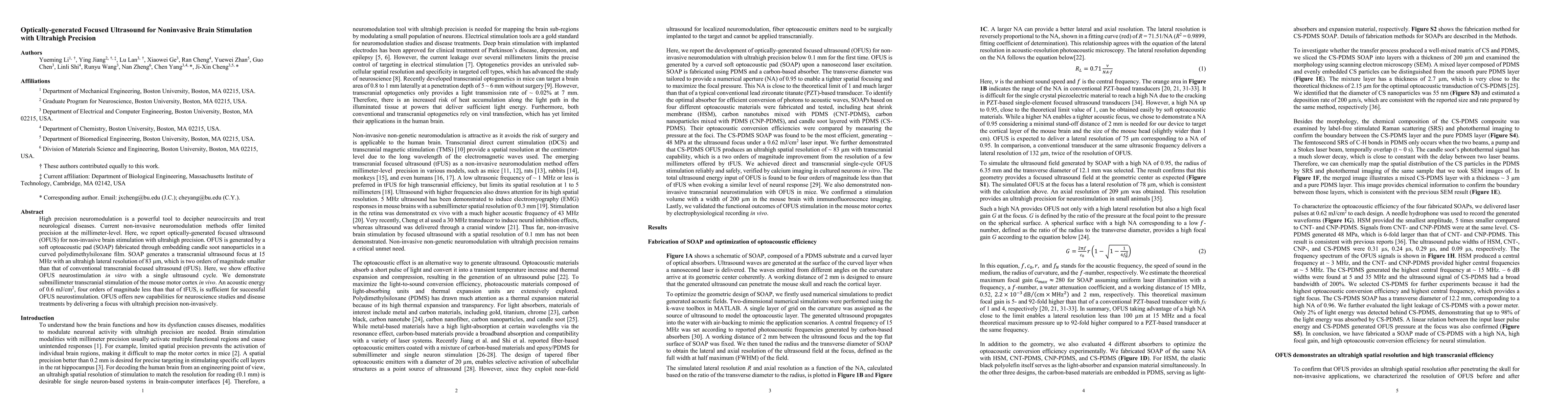
High precision neuromodulation is a powerful tool to decipher neurocircuits and treat neurological diseases. Current non-invasive neuromodulation methods offer limited precision at the millimeter le...
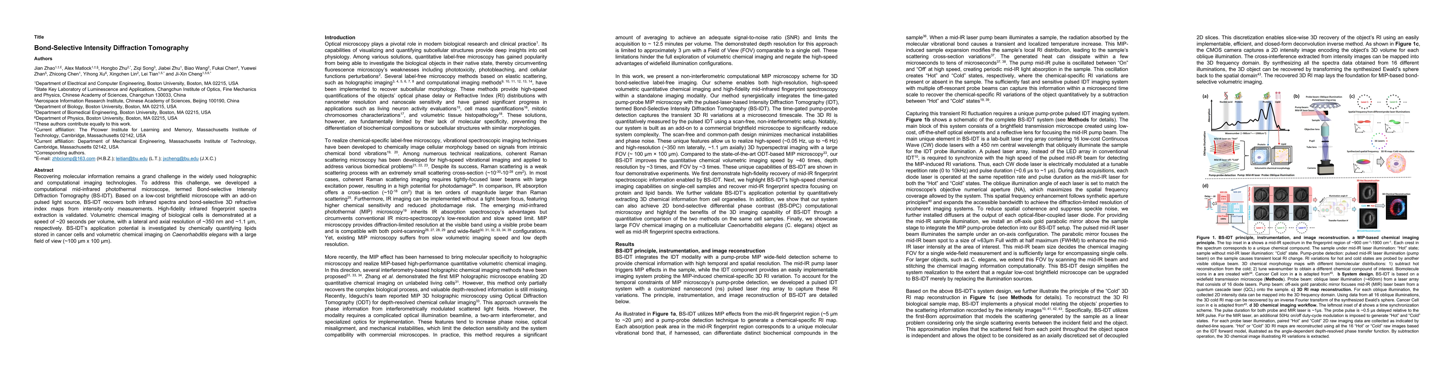
Recovering molecular information remains a grand challenge in the widely used holographic and computational imaging technologies. To address this challenge, we developed a computational mid-infrared...
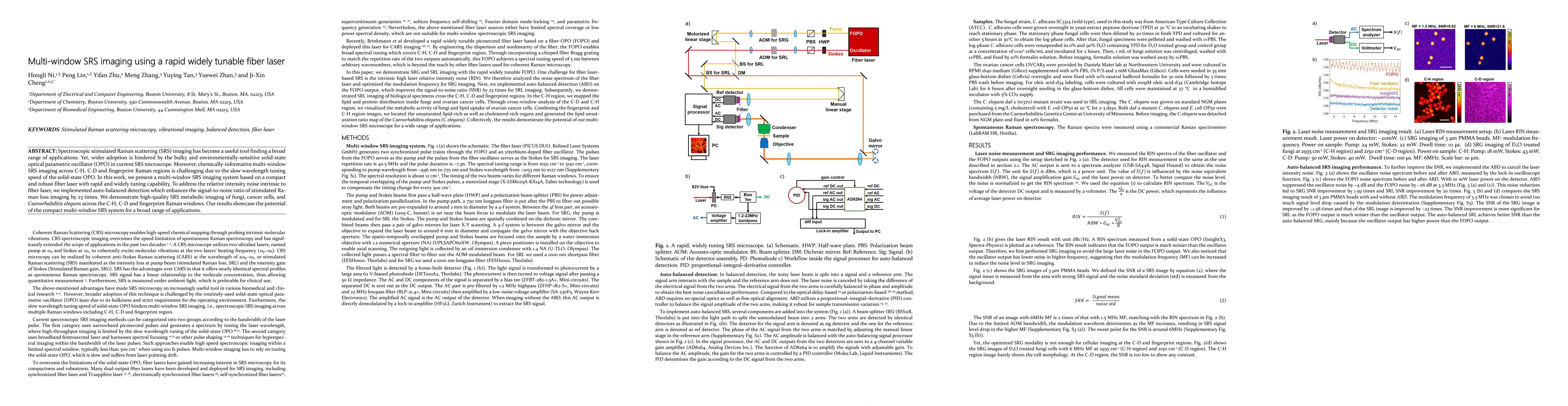
Spectroscopic stimulated Raman scattering (SRS) imaging has become a useful tool finding a broad range of applications. Yet, wider adoption is hindered by the bulky and environmentally-sensitive sol...
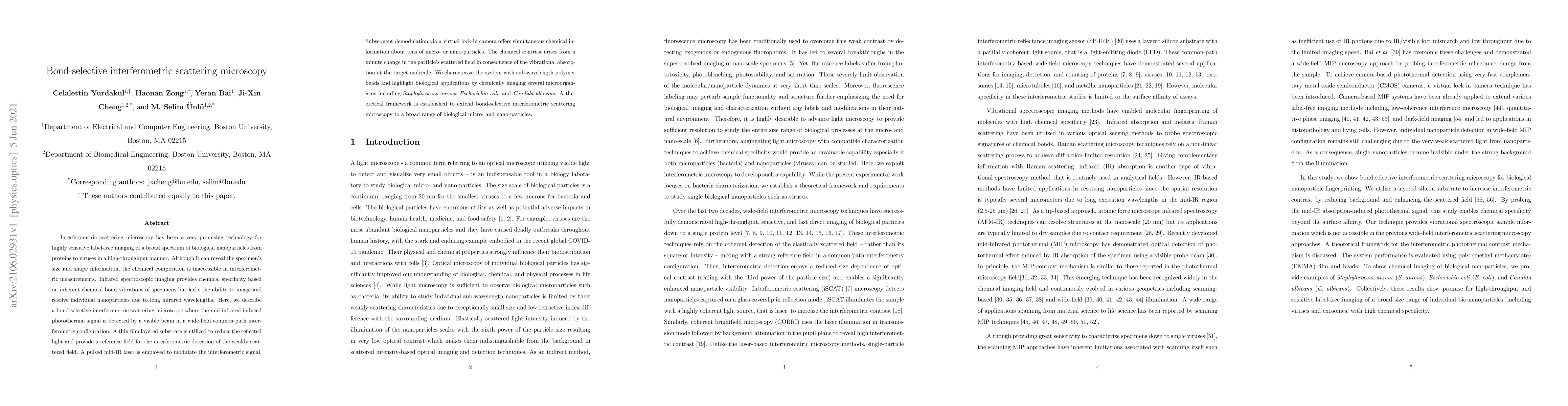
Interferometric scattering microscopy has been a very promising technology for highly sensitive label-free imaging of a broad spectrum of biological nanoparticles from proteins to viruses in a high-...
Mid-infrared photothermal (MIP) microscopy has been a promising label-free chemical imaging technique for functional characterization of specimens owing to its enhanced spatial resolution and high s...
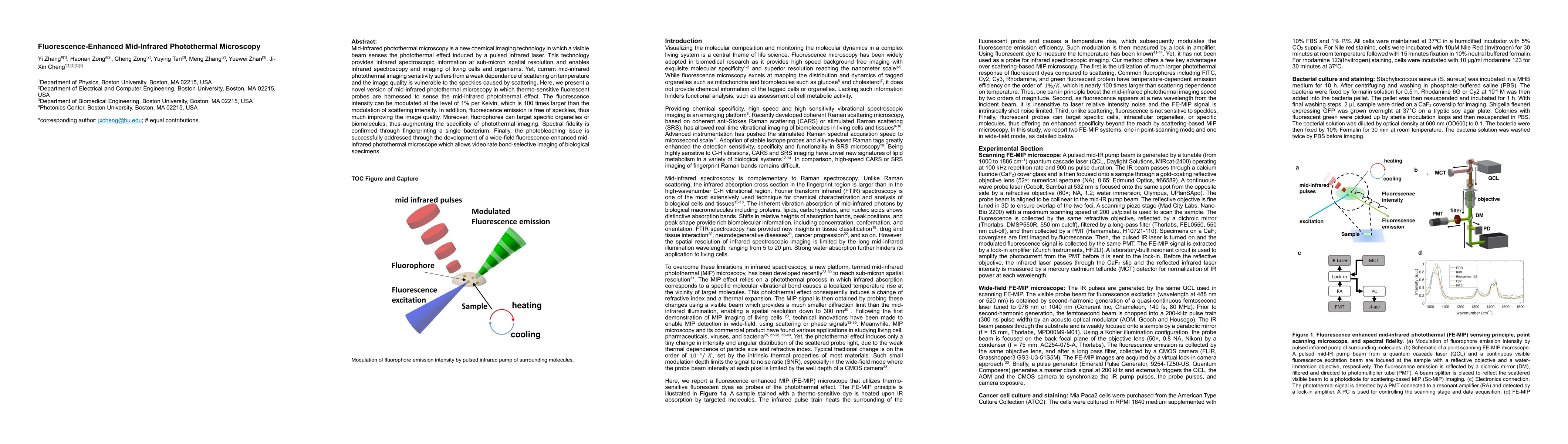
Mid-infrared photothermal microscopy is a new chemical imaging technology in which a visible beam senses the photothermal effect induced by a pulsed infrared laser. This technology provides infrared...
As an emerging technology, transcranial focused ultrasound has been demonstrated to successfully evoke motor responses in mice, rabbits, and sensory/motor responses in humans. Yet, the spatial resol...
Label-free vibrational imaging by stimulated Raman scattering (SRS) provides unprecedented insight into real-time chemical distributions in living systems. Specifically, SRS in the fingerprint regio...
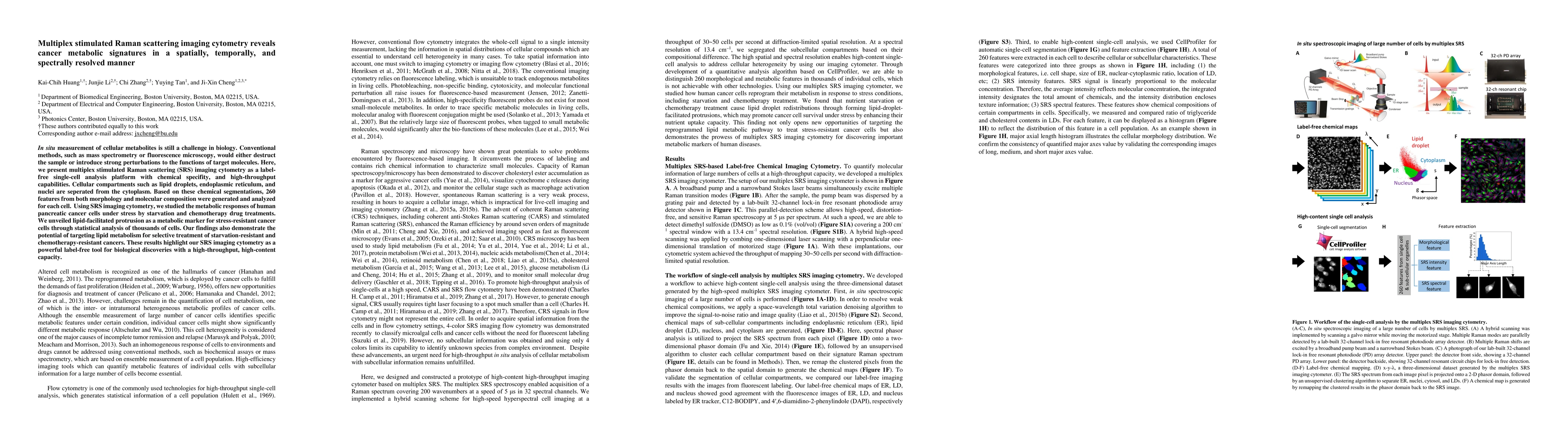
In situ measurement of cellular metabolites is still a challenge in biology. Conventional methods, such as mass spectrometry or fluorescence microscopy, would either destruct the sample or introduce...
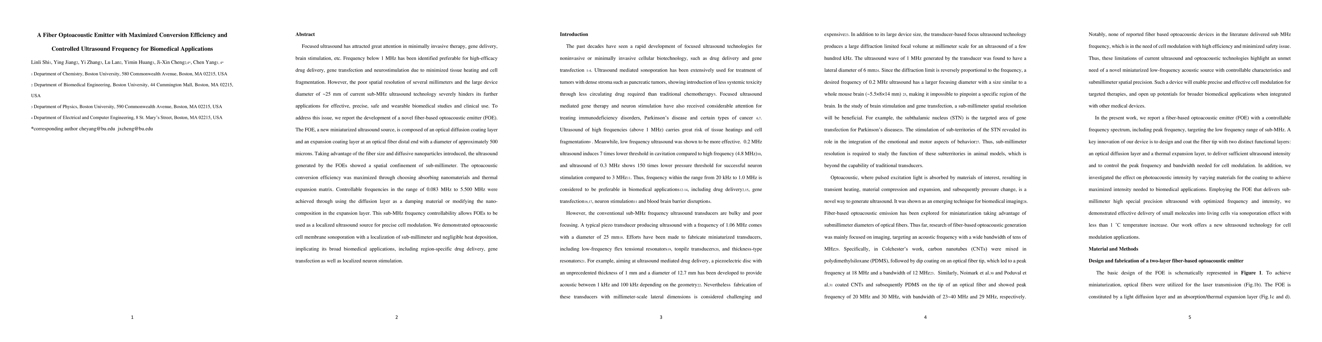
Focused ultrasound has attracted great attention in minimally invasive therapy, gene delivery, brain stimulation, etc. Frequency below 1 MHz has been identified preferable for high-efficacy drug del...
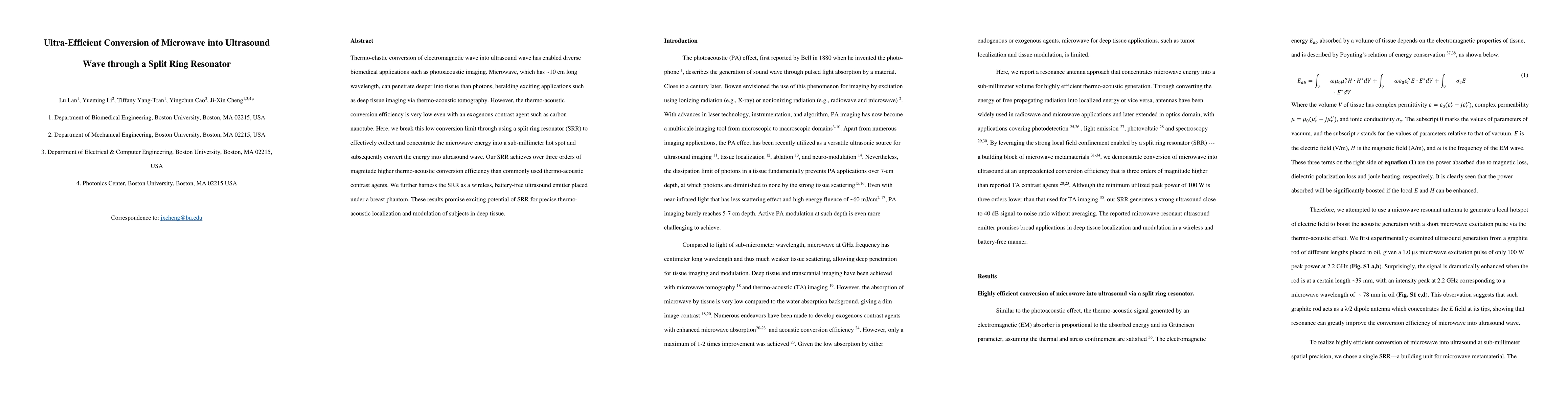
Thermo-elastic conversion of electromagnetic wave into ultrasound wave has enabled diverse biomedical applications such as photoacoustic imaging. Microwave, which has ~10 cm long wavelength, can pen...
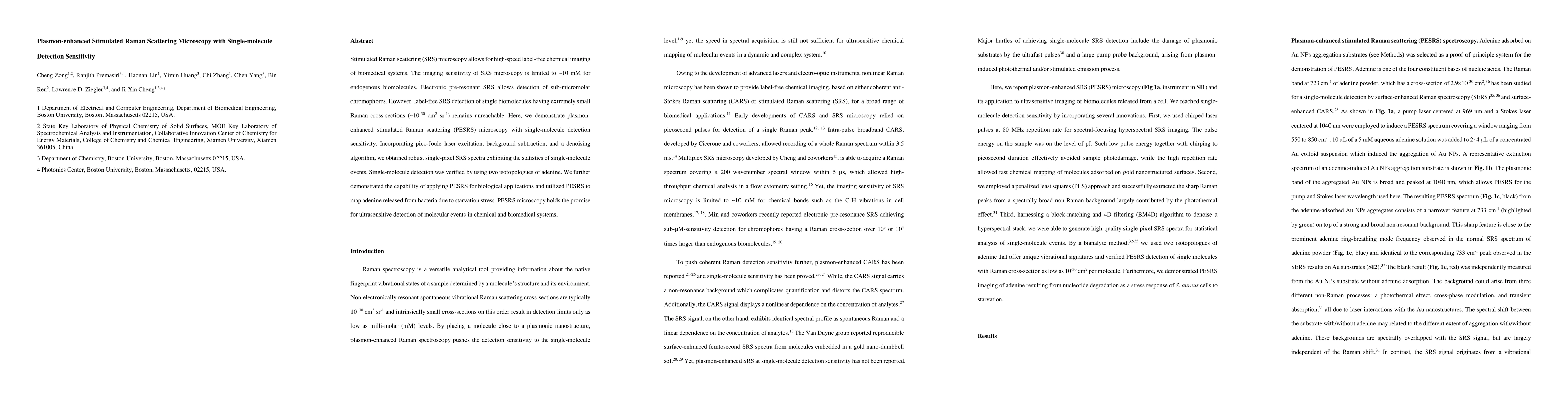
Stimulated Raman scattering (SRS) microscopy allows for high-speed label-free chemical imaging of biomedical systems. The imaging sensitivity of SRS microscopy is limited to ~10 mM for endogenous bi...
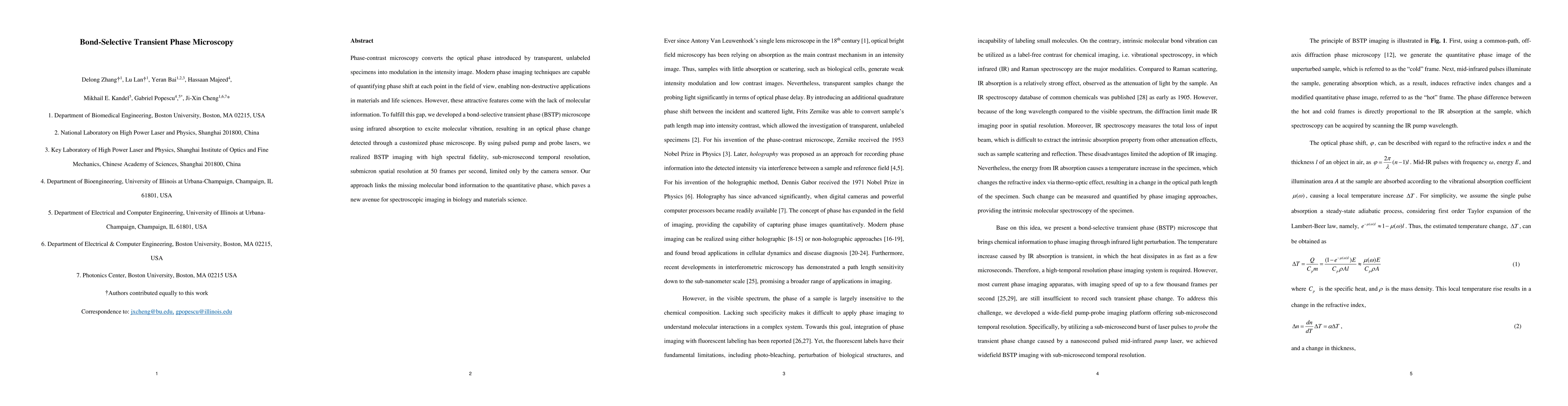
Phase-contrast microscopy converts the optical phase introduced by transparent, unlabeled specimens into modulation in the intensity image. Modern phase imaging techniques are capable of quantifying...
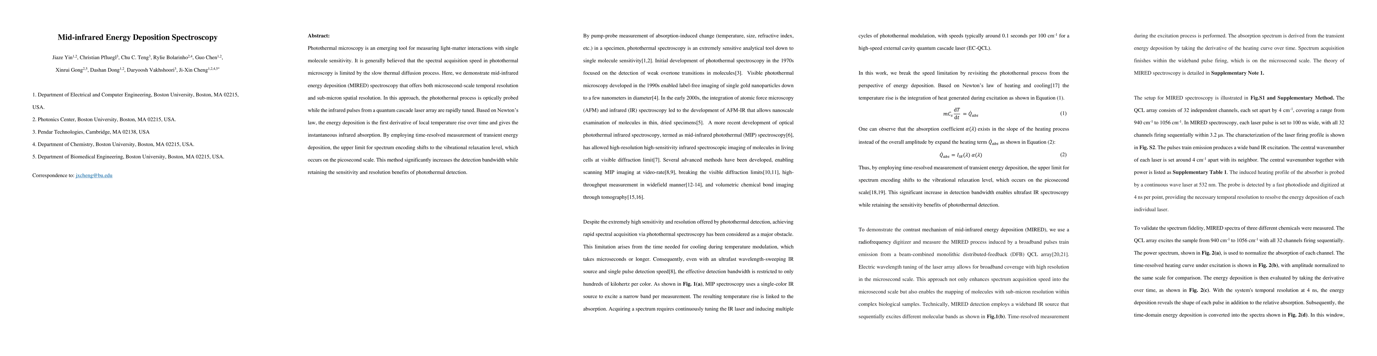
Photothermal microscopy is an emerging tool for measuring light-matter interactions with single-molecule sensitivity. It is generally believed that the spectral acquisition speed in photothermal micro...
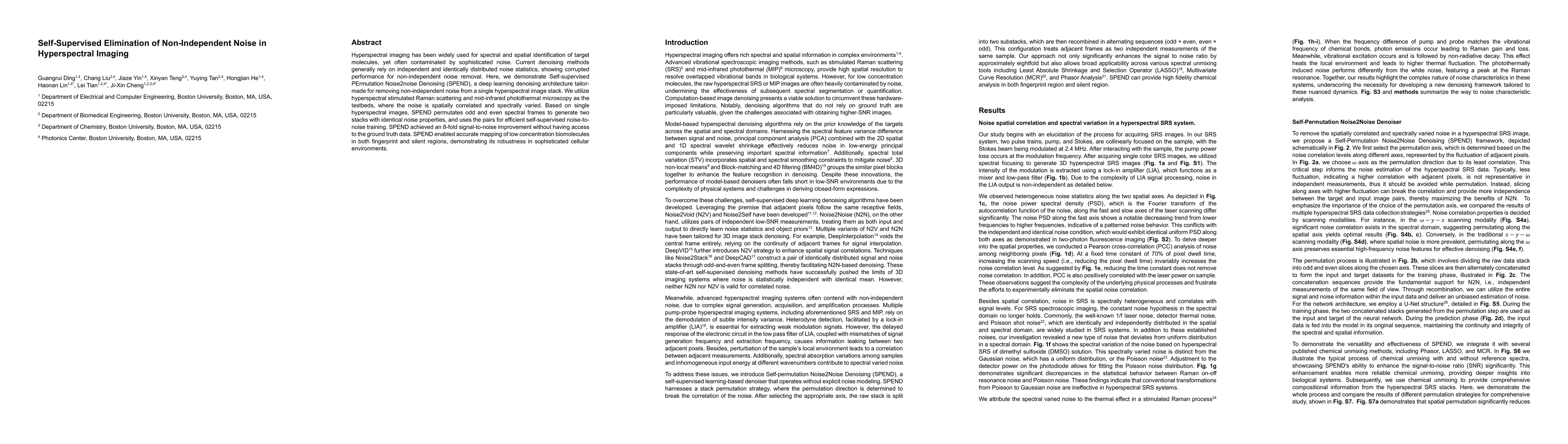
Hyperspectral imaging has been widely used for spectral and spatial identification of target molecules, yet often contaminated by sophisticated noise. Current denoising methods generally rely on indep...
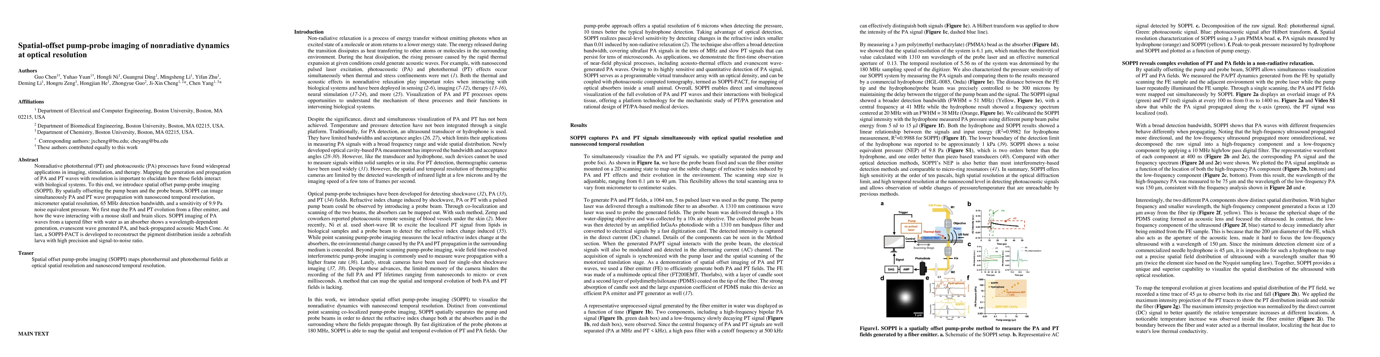
Nonradiative photothermal (PT) and photoacoustic (PA) processes have found widespread applications in imaging, stimulation, and therapy. Mapping the generation and propagation of PA and PT waves with ...
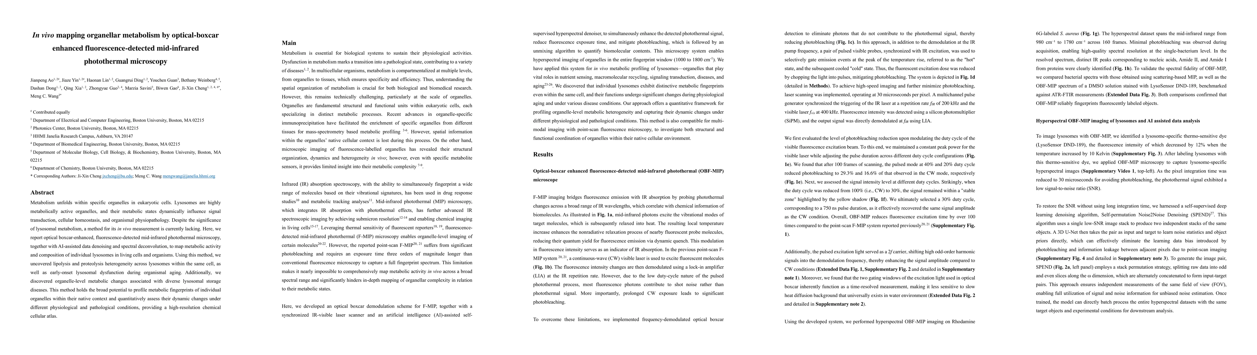
Metabolism unfolds within specific organelles in eukaryotic cells. Lysosomes are highly metabolically active organelles, and their metabolic states dynamically influence signal transduction, cellular ...
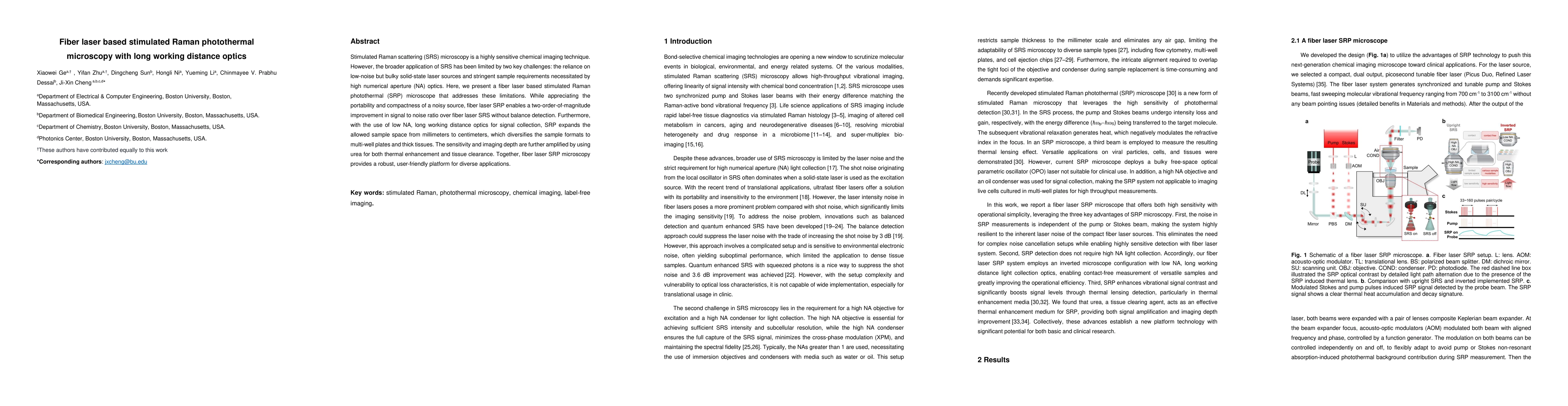
Stimulated Raman scattering (SRS) microscopy is a highly sensitive chemical imaging technique. However, the broader application of SRS has been limited by two key challenges: the reliance on low-noise...
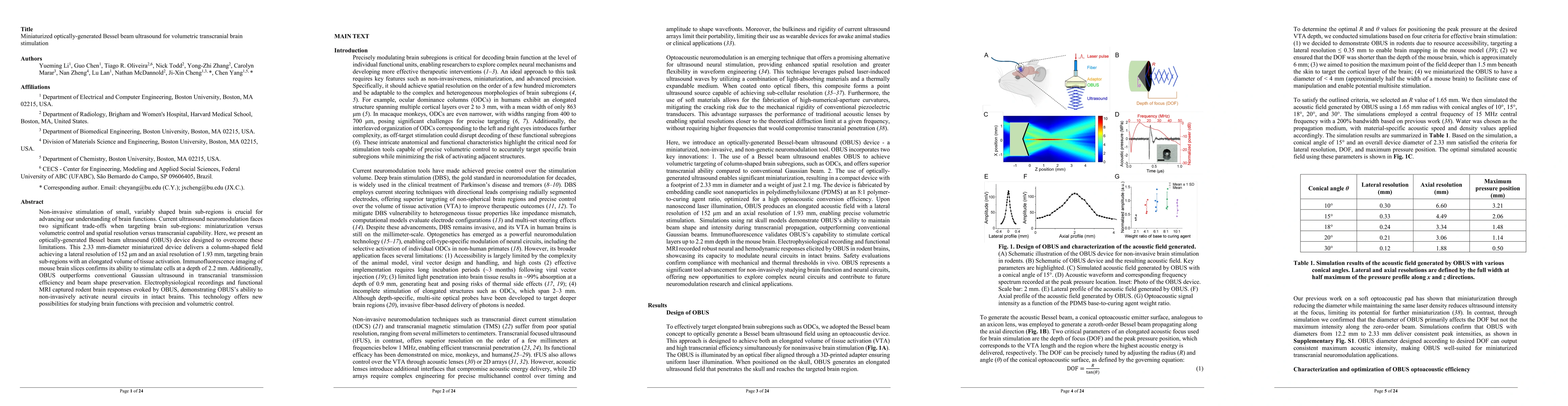
Non-invasive stimulation of small, variably shaped brain sub-regions is crucial for advancing our understanding of brain functions. Current ultrasound neuromodulation faces two significant trade-offs ...
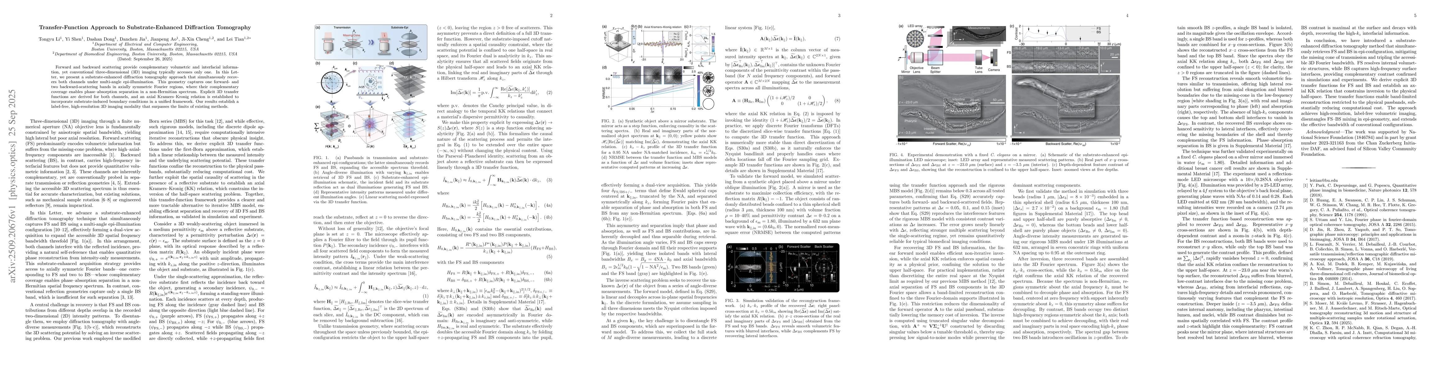
Forward and backward scattering provide complementary volumetric and interfacial information, yet conventional three-dimensional (3D) imaging typically accesses only one. In this Letter, we present a ...
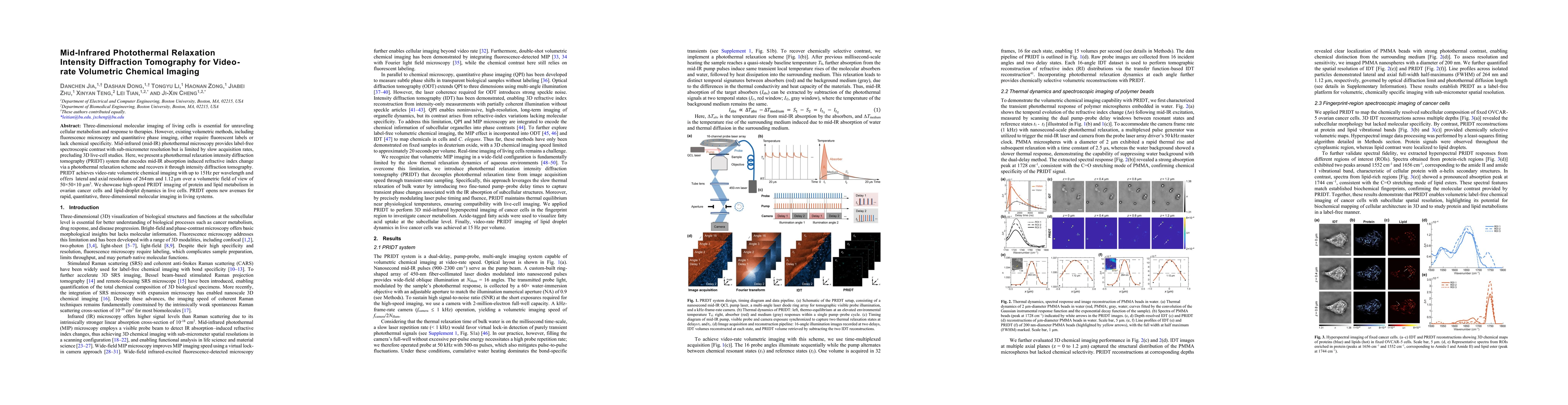
Three-dimensional molecular imaging of living cells is essential for unraveling cellular metabolism and response to therapies. However, existing volumetric methods, including fluorescence microscopy a...
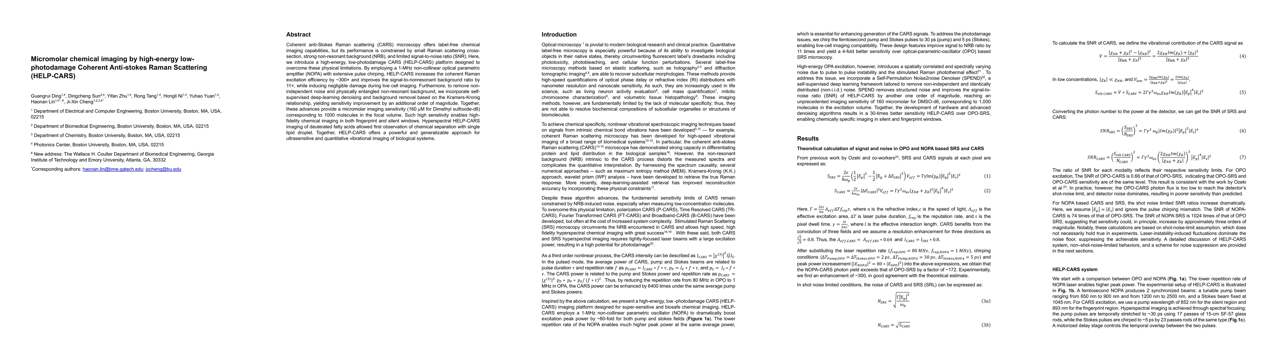
Coherent anti-Stokes Raman scattering (CARS) microscopy offers label-free chemical imaging capabilities, but its performance is constrained by small Raman scattering cross-section, strong non-resonant...
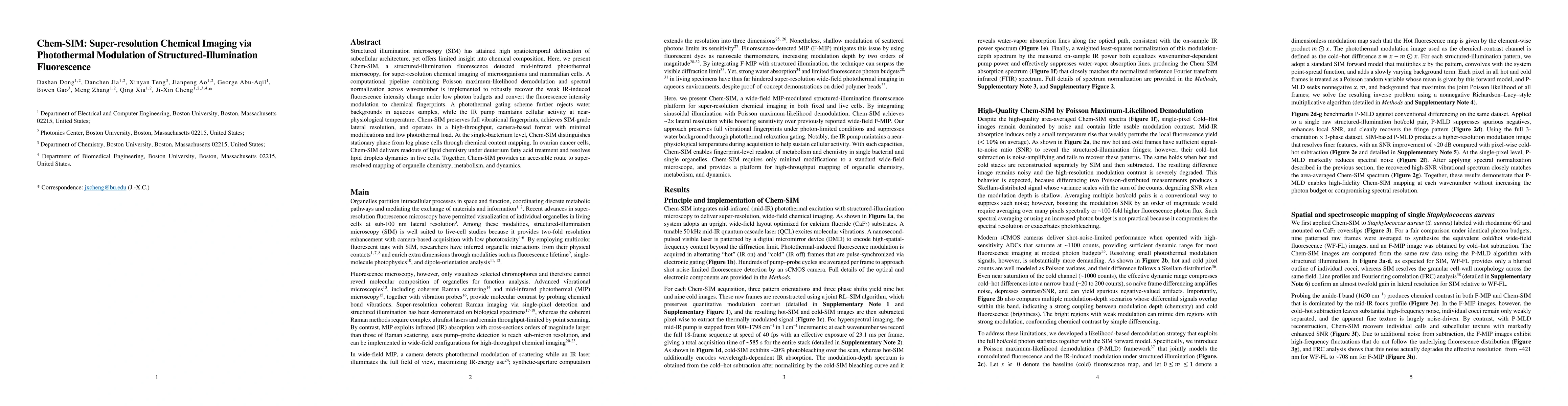
Structured illumination microscopy (SIM) has attained high spatiotemporal delineation of subcellular architecture, yet offers limited insight into chemical composition. Here, we present Chem-SIM, a st...
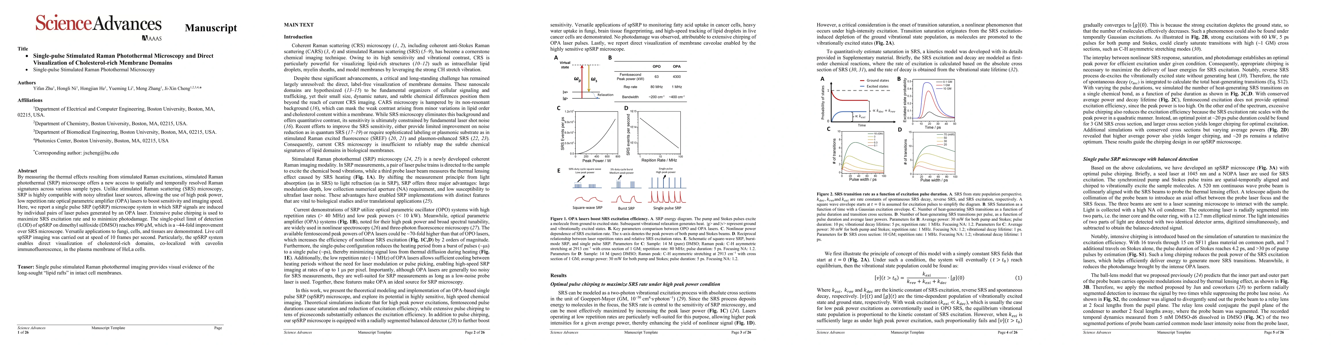
By measuring the thermal effects resulting from stimulated Raman excitations, stimulated Raman photothermal (SRP) microscope offers a new access to spatially and temporally resolved Raman signatures a...
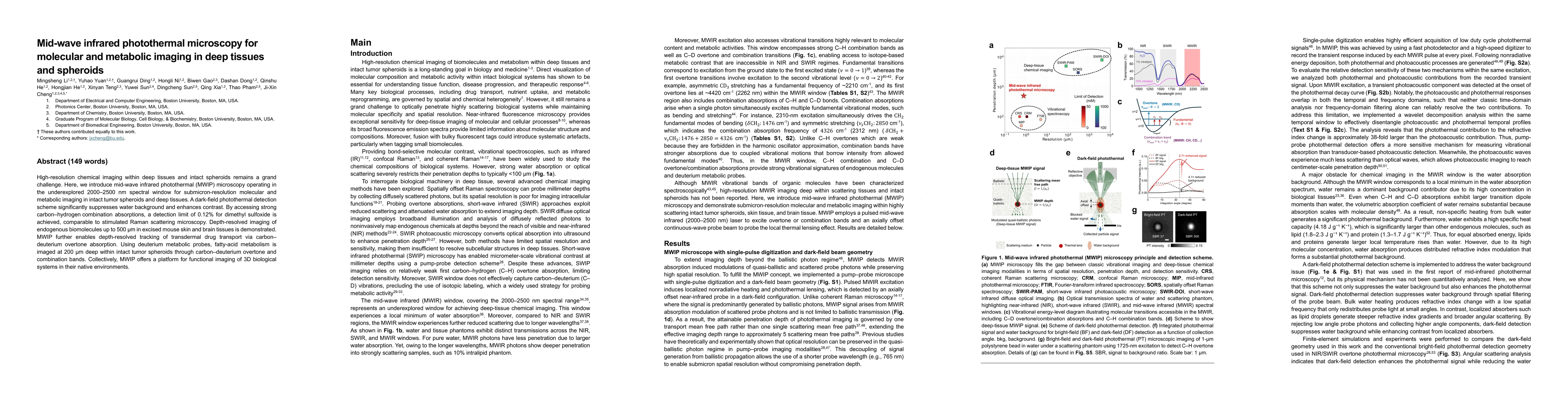
High-resolution chemical imaging within deep tissues and intact spheroids remains a grand challenge. Here, we introduce mid-wave infrared photothermal (MWIP) microscopy operating in the underexplored ...
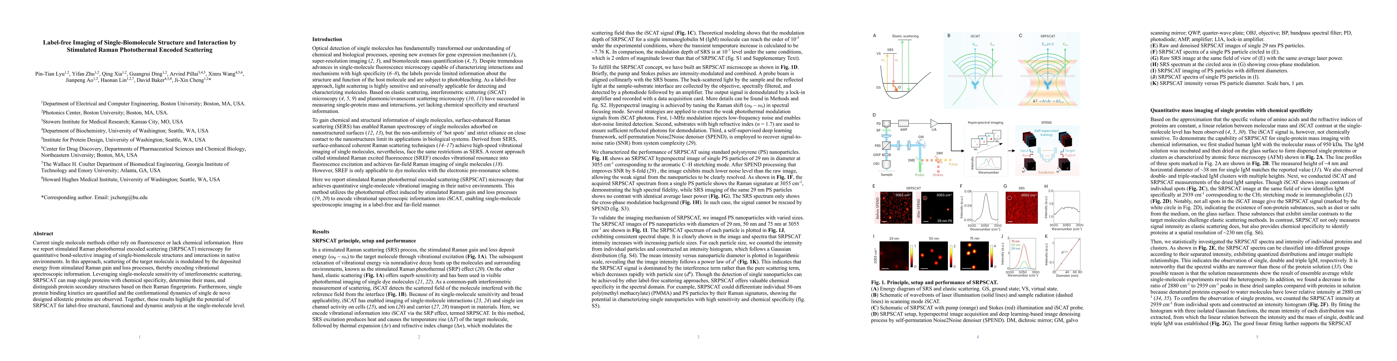
Current single molecule methods either rely on fluorescence or lack chemical information. Here we report stimulated Raman photothermal encoded scattering (SRPSCAT) microscopy for quantitative bond-sel...
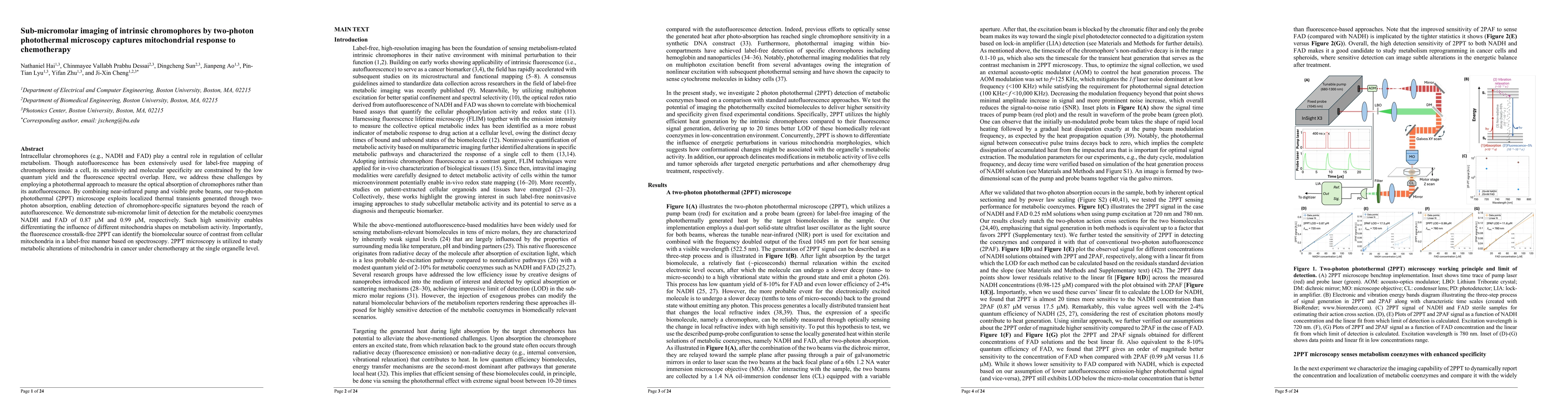
Intracellular chromophores (e.g., NADH and FAD) play a central role in regulation of cellular metabolism. Though autofluorescence has been extensively used for label-free mapping of chromophores insid...