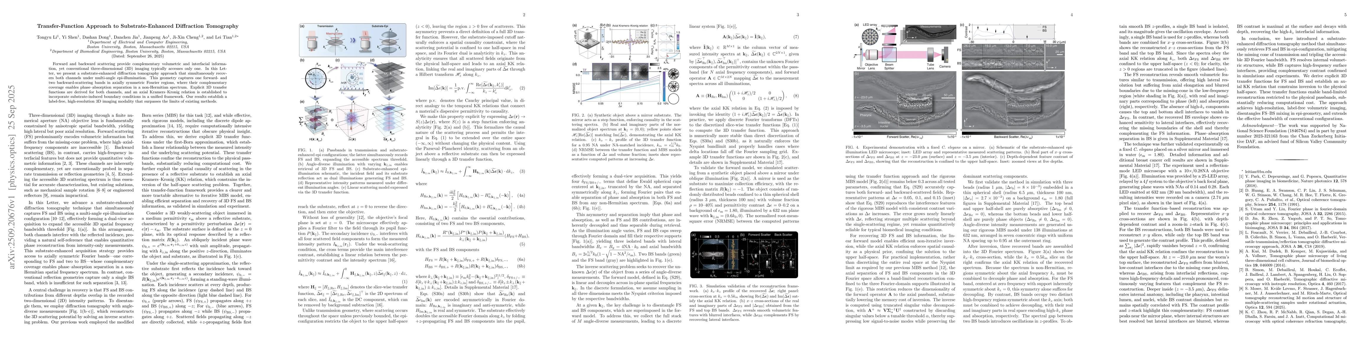
Summary

Forward and backward scattering provide complementary volumetric and interfacial information, yet conventional three-dimensional (3D) imaging typically accesses only one. In this Letter, we present a substrate-enhanced diffraction tomography approach that simultaneously recovers both channels under multi-angle epi-illumination.

AI Key Findings

Generated Sep 26, 2025

Methodology

The research employs a combination of advanced optical imaging techniques and computational reconstruction algorithms to analyze 3D phase and absorption information from scattered light. It utilizes a band-limited reconstruction approach with a transfer function-based method to decouple phase and absorption components from forward and backward scattering data.

Key Results

  • Successful decoupling of 3D phase and absorption information using both forward and backward scattering data
  • Reconstruction of complex biological structures like C. elegans and breast cancer cells with high spatial resolution
  • Development of an efficient inversion algorithm that enables rapid 3D reconstruction of multiple measurement blocks

Significance

This research advances optical imaging by enabling non-invasive 3D characterization of biological samples with high resolution. The methodology could have applications in biomedical imaging, material science, and non-destructive testing of complex structures.

Technical Contribution

The technical contribution lies in the development of a band-limited reconstruction algorithm that effectively separates phase and absorption information from forward and backward scattering data using transfer function analysis.

Novelty

This work introduces a novel approach to 3D optical reconstruction by combining transfer function analysis with band-limited reconstruction, enabling simultaneous recovery of both phase and absorption information from scattered light with improved efficiency compared to existing methods.

Limitations

  • Requires careful calibration of the inversion operator for different measurement configurations
  • Limited depth resolution compared to traditional microscopy techniques

Future Work

  • Integration with machine learning for automated parameter optimization
  • Extension to higher resolution and larger field-of-view applications
  • Development of real-time reconstruction systems for clinical applications

Paper Details

PDF Preview

Download PDF

Comments (0)

This website uses cookies to ensure you get the best experience. Learn more