Millimeter-deep micron-resolution vibrational imaging by shortwave infrared photothermal microscopy
Publication
Metrics
AI Quick Summary
Shortwave infrared photothermal microscopy (SWIP) enables millimeter-deep vibrational imaging with sub-micron resolution, detecting chemical contrasts in biological tissues and nanoparticles. This technique provides enhanced sensitivity, resolving cellular structures in liver, skin, brain, and breast tissues, promising broad applications in life sciences and clinical diagnostics.
Paper Preview
Abstract
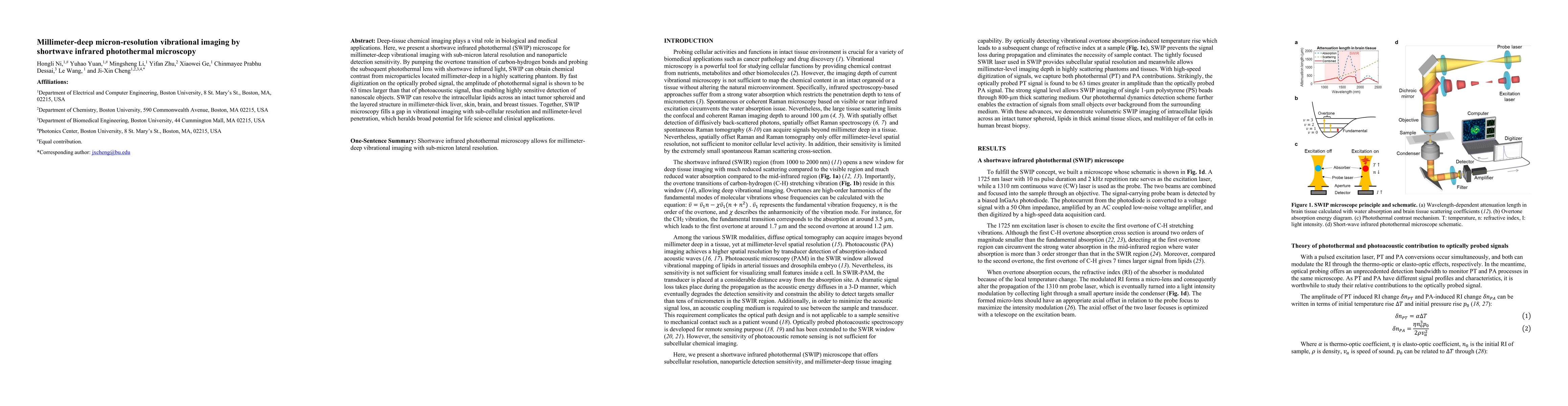
Deep-tissue chemical imaging plays a vital role in biological and medical applications. Here, we present a shortwave infrared photothermal (SWIP) microscope for millimeter-deep vibrational imaging with sub-micron lateral resolution and nanoparticle detection sensitivity. By pumping the overtone transition of carbon-hydrogen bonds and probing the subsequent photothermal lens with shortwave infrared light, SWIP can obtain chemical contrast from microparticles located millimeter-deep in a highly scattering phantom. By fast digitization on the optically probed signal, the amplitude of photothermal signal is shown to be 63 times larger than that of photoacoustic signal, thus enabling highly sensitive detection of nanoscale objects. SWIP can resolve the intracellular lipids across an intact tumor spheroid and the layered structure in millimeter-thick liver, skin, brain, and breast tissues. Together, SWIP microscopy fills a gap in vibrational imaging with sub-cellular resolution and millimeter-level penetration, which heralds broad potential for life science and clinical applications.
AI Key Findings
Get AI-generated insights about this paper's methodology, results, significance, and more — seven facets brought into focus.
Impact
Paper Details
Authors
PDF Preview
Key Terms
Citation Network
Current paper (gray), citations (green), references (blue)
Display is limited for performance on very large graphs.
Discussion 0