Quick Summary:

This paper presents a two-photon light-sheet microscopy setup that significantly enhances the imaging speed for whole-brain functional imaging in zebrafish larvae, achieving a 5 Hz acquisition rate while minimizing laser intensity. The method is used to study seizures, revealing caudo-rostral ictal waves for the first time.

Summary

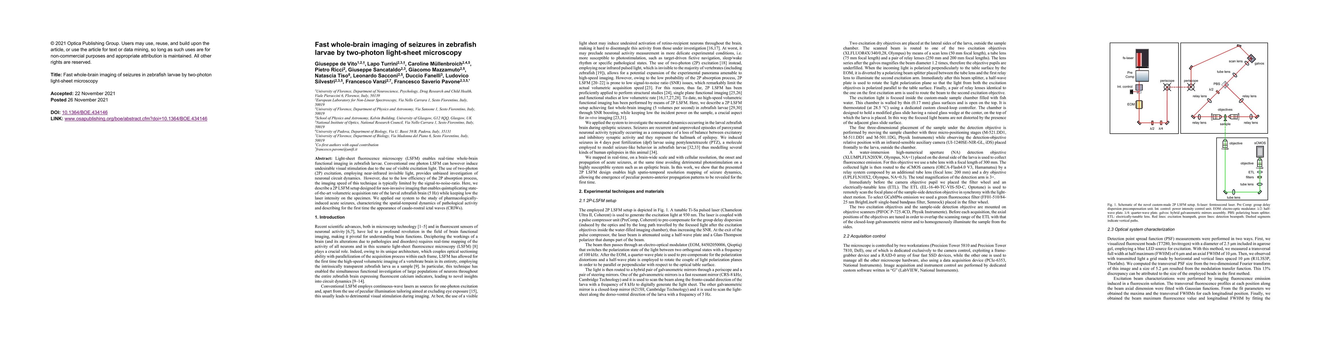
Light-sheet fluorescence microscopy (LSFM) enables real-time whole-brain functional imaging in zebrafish larvae. Conventional one photon LSFM can however induce undesirable visual stimulation due to the use of visible excitation light. The use of two-photon (2P) excitation, employing near-infrared invisible light, provides unbiased investigation of neuronal circuit dynamics. However, due to the low efficiency of the 2P absorption process, the imaging speed of this technique is typically limited by the signal-to-noise-ratio. Here, we describe a 2P LSFM setup designed for non-invasive imaging that enables quintuplicating state-of-the-art volumetric acquisition rate of the larval zebrafish brain (5 Hz) while keeping low the laser intensity on the specimen. We applied our system to the study of pharmacologically-induced acute seizures, characterizing the spatial-temporal dynamics of pathological activity and describing for the first time the appearance of caudo-rostral ictal waves (CRIWs).

AI Key Findings

Get AI-generated insights about this paper's methodology, results, and significance.

Paper Details

PDF Preview

Download PDF

Key Terms

brain (0.242)
imaging (0.231)
excitation (0.195)
photon (0.193)
light (0.187)
enables (0.179)
fluorescence (0.153)
sheet (0.150)
hz (0.149)
unbiased (0.146)
noise ratio (0.145)
near infrared (0.143)
dynamics (0.141)
signal noise (0.140)
characterizing (0.140)
keeping (0.139)
acquisition (0.137)
circuit (0.136)
microscopy (0.133)
setup (0.132)

Citation Network

Current paper (gray), citations (green), references (blue)

Current Paper
Citations
References
Click to view
Title Authors Year Actions
Fluorescence spatial anisotropy of emission dipoles in an orthogonal imaging system
Yahui Song, Xin Xu, Jixiang Wang, Lina Xing, Xin Zhang, Guohua Shi, Hong Ye
2025
View Paper
Near infrared‐II light‐sheet microscopy: Basic principle, intellectualization, and medical application
Yuan Li, Siyu Ao, Tianyu Zhu, Rong Zhao, Hao Yang, Qizhong Wang, Xiaoyu Mu, Hao Wang, Pengfei Liu, Xiaodong Zhang
2024
View Paper
Epilepsy insights revealed by intravital functional optical imaging
Matthew A. Stern, Raymond Dingledine, Robert E. Gross, Ken Berglund
2024
View Paper
More than double the fun with two-photon excitation microscopy
Peter Luu, Scott E. Fraser, Falk Schneider
2024
View Paper
Two-photon all-optical electrophysiology for the dissection of larval zebrafish brain functional connectivity
L. Turrini, Pietro Ricci, Michele Sorelli, Giuseppe de Vito, Marco Marchetti, F. Vanzi, Francesco S. Pavone
2024
View Paper
Introduction to the Optics and the Brain 2023 feature issue
Adam Q. Bauer, E. A. Gibson, Hui Wang, Vivek Srinivasan
2024
View Paper
Open-top Bessel beam two-photon light sheet microscopy for three-dimensional pathology
W. Park, Jieun Yun, Jinho Shin, B. Oh, Gilsuk Yoon, Seung-Mo Hong, Ki Hean Kim
2023
View Paper
Small fish, big discoveries: zebrafish shed light on microbial biomarkers for neuro-immune-cardiovascular health
Hemaa Sree Kumar, Alexander S. Wisner, Jeanine M. Refsnider, C. Martyniuk, J. Zubcevic
2023
View Paper
Imaging Approaches to Investigate Pathophysiological Mechanisms of Brain Disease in Zebrafish
L. Turrini, Lorenzo Roschi, Giuseppe de Vito, F. Pavone, F. Vanzi
2023
View Paper
Enhanced neuroimaging with a calcium sensor in ex-vivo Drosophila melanogaster brains using closed-loop adaptive optics light-sheet fluorescence microscopy
A. Hubert, Georges Farkouh, F. Harms, Cynthia Veilly, S. Imperato, Mathias Mercier, V. Loriette, F. Rouyer, A. Fragola
2023
View Paper
Zebrafish as an Innovative Tool for Epilepsy Modeling: State of the Art and Potential Future Directions
M. d’Amora, A. Galgani, M. Marchese, F. Tantussi, U. Faraguna, F. De Angelis, F. Giorgi
2023
View Paper
Brain-wide functional imaging to highlight differences between the diurnal and nocturnal neuronal activity in zebrafish larvae
G. Vito, L. Turrini, C. Fornetto, Elena Trabalzini, Pietro Ricci, D. Fanelli, F. Vanzi, F. Pavone
2023
View Paper
Fast multi-photon brain-wide volumetric imaging and photostimulation
L. Turrini, Pietro Ricci, Giuseppe de Vito, Michele Sorelli, M. Marchetti, F. Vanzi, L. Silvestri, F. Pavone
2023
View Paper
Enhanced neuroimaging with a calcium sensor in the live adult Drosophila Melanogaster brain using closed-loop adaptive optics light-sheet microscopy
A. Hubert, Georges Farkouh, F. Harms, Cynthia Veilly, S. Imperato, Mathias Mercier, V. Loriette, F. Rouyer, A. Fragola
2023
View Paper
Quantity as a Fish Views It: Behavior and Neurobiology
A. Messina, Davide Potrich, Matilde Perrino, Eva Sheardown, M. E. Miletto Petrazzini, Peter Luu, Anna Nadtochiy, T. Truong, V. A. Sovrano, S. Fraser, C. Brennan, G. Vallortigara
2022
View Paper
Extended depth-of-field light-sheet microscopy improves imaging of large volumes at high numerical aperture
Kevin Keomanee-Dizon, Matt Jones, Peter Luu, S. Fraser, T. Truong
2022
View Paper
Multimodal Characterization of Seizures in Zebrafish Larvae
L. Turrini, Michele Sorelli, Giuseppe de Vito, C. Credi, N. Tiso, F. Vanzi, F. Pavone
2022
View Paper
Real time large scale $\textit{in vivo}$ observations by light-sheet microscopy reveal intrinsic synchrony, plasticity and growth cone dynamics of midline crossing axons at the ventral floor plate of the zebrafish spinal cord
Søren S L Andersen
2021
View Paper
Optical volumetric brain imaging: speed, depth, and resolution enhancement
Shih-Huan Huang, N. Irawati, Yu-Feng Chien, Jyun-Yi Lin, Yu-Hsuan Tsai, Po-Yuan Wang, L. Chu, Meng-Lin Li, A. Chiang, K. Tsia, S. Chu
2021
View Paper

Comments (0)

This website uses cookies to ensure you get the best experience. Learn more