Polarization effects on fluorescence emission of zebrafish neurons using light-sheet microscopy
Publication
Metrics
AI Quick Summary
This paper investigates the polarization effects on fluorescence emission in zebrafish neurons using light-sheet microscopy, revealing a 40% higher emission when the polarization is orthogonal to the illumination and detection axes. The enhanced signals predominantly originate from nerve cells, improving contrast and resolution for observing organism structures.
Paper Preview
Abstract
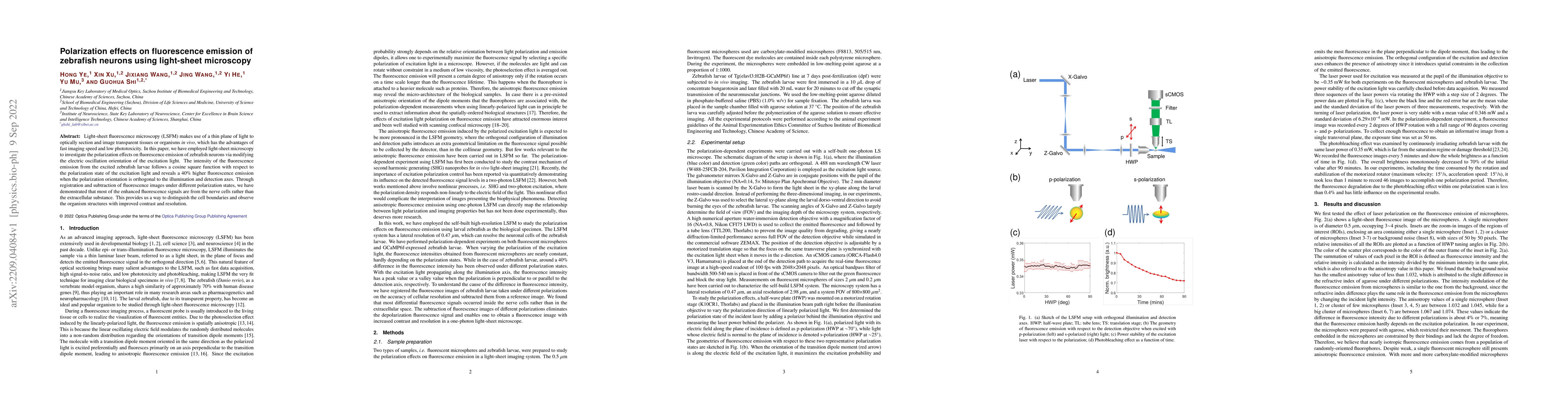
Light-sheet fluorescence microscopy (LSFM) makes use of a thin plane of light to optically section and image transparent tissues or organisms {\it{in vivo}}, which has the advantages of fast imaging speed and low phototoxicity. In this paper, we have employed light-sheet microscopy to investigate the polarization effects on fluorescence emission of zebrafish neurons via modifying the electric oscillation orientation of the excitation light. The intensity of the fluorescence emission from the excited zebrafish larvae follows a cosine square function with respect to the polarization state of the excitation light and reveals a 40$\%$ higher fluorescence emission when the polarization orientation is orthogonal to the illumination and detection axes. Through registration and subtraction of fluorescence images under different polarization states, we have demonstrated that most of the enhanced fluorescence signals are from the nerve cells rather than the extracellular substance. This provides us a way to distinguish the cell boundaries and observe the organism structures with improved contrast and resolution.
AI Key Findings
Get AI-generated insights about this paper's methodology, results, significance, and more — seven facets brought into focus.
Impact
Paper Details
Authors
PDF Preview
Key Terms
Citation Network
Current paper (gray), citations (green), references (blue)
Display is limited for performance on very large graphs.
Discussion 0