Publication
Metrics
Paper Preview
Abstract
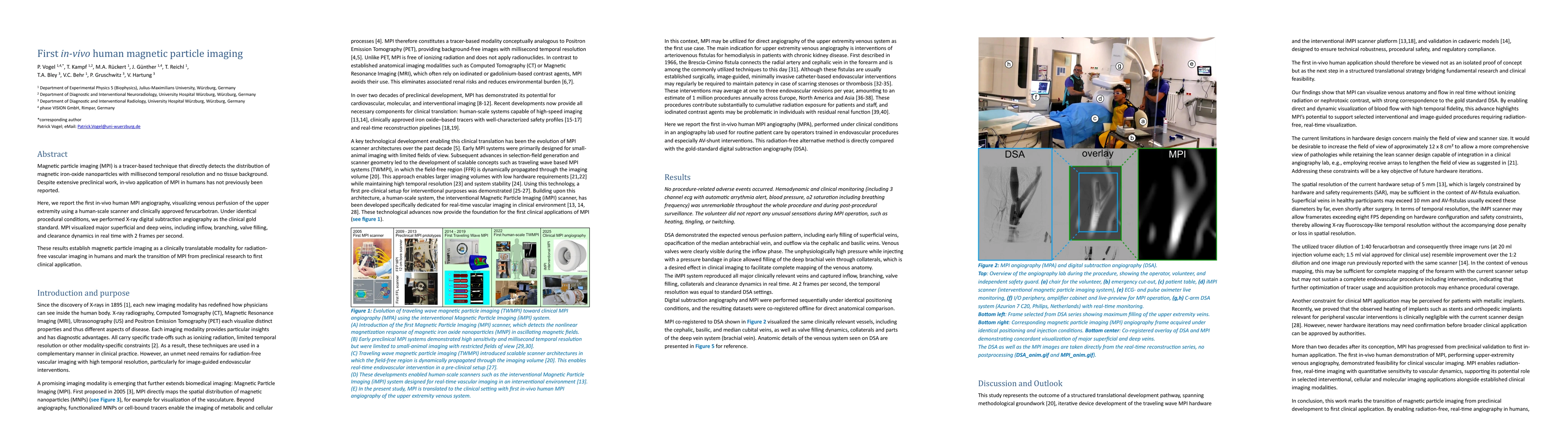
Magnetic particle imaging (MPI) is a tracer-based technique that directly detects the distribution of magnetic iron-oxide nanoparticles with millisecond temporal resolution and no tissue background. Despite extensive preclinical work, in-vivo application of MPI in humans has not previously been reported. Here, we report the first in-vivo human MPI angiography, visualizing venous perfusion of the upper extremity using a human-scale scanner and clinically approved ferucarbotran. Under identical procedural conditions, we performed X-ray digital subtraction angiography as the clinical gold standard. MPI visualized major superficial and deep veins, including inflow, branching, valve filling, and clearance dynamics in real time with 2 frames per second. These results establish magnetic particle imaging as a clinically translatable modality for radiation-free vascular imaging in humans and mark the transition of MPI from preclinical research to first clinical application.
AI Key Findings
Get AI-generated insights about this paper's methodology, results, significance, and more — seven facets brought into focus.
Discussion 0