First Indirect X-Ray Imaging Tests With An 88-mm Diameter Single Crystal
Publication
Metrics
AI Quick Summary
This paper reports the first indirect x-ray imaging tests using an 88-mm diameter YAG:Ce single crystal, achieving a 10.5 micron point-spread-function (PSF) resolution, significantly better than standard phosphors. The crystal was bonded to a fiber optic plate for coupling to a camera, demonstrating potential for future x-ray diffraction experiments.
Paper Preview
Abstract
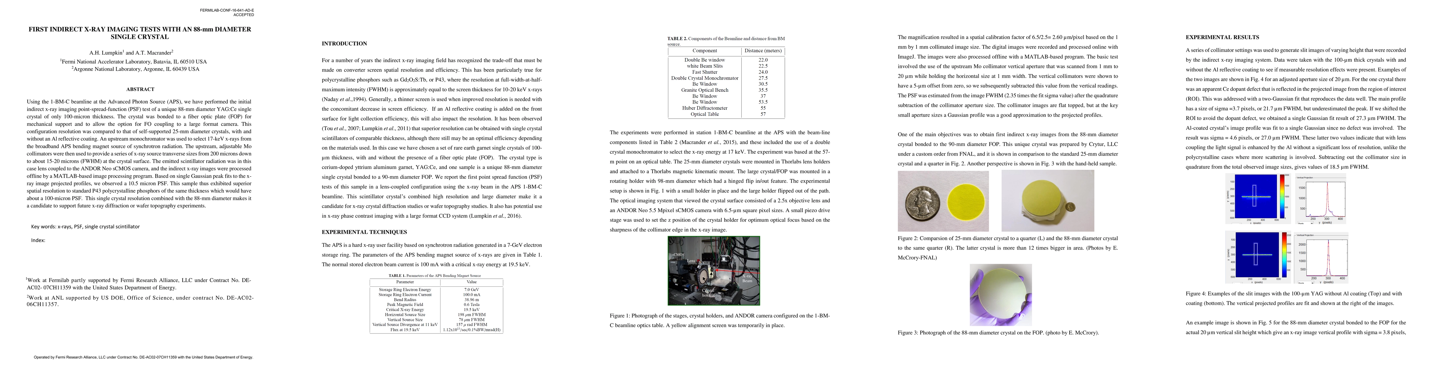
Using the 1-BM-C beamline at the Advanced Photon Source (APS), we have performed the initial indirect x-ray imaging point-spread-function (PSF) test of a unique 88-mm diameter YAG:Ce single crystal of only 100-micron thickness. The crystal was bonded to a fiber optic plate (FOP) for mechanical support and to allow the option for FO coupling to a large format camera. This configuration resolution was compared to that of self-supported 25-mm diameter crystals, with and without an Al reflective coating. An upstream monochromator was used to select 17-keV x-rays from the broadband APS bending magnet source of synchrotron radiation. The upstream, adjustable Mo collimators were then used to provide a series of x-ray source transverse sizes from 200 microns down to about 15-20 microns (FWHM) at the crystal surface. The emitted scintillator radiation was in this case lens coupled to the ANDOR Neo sCMOS camera, and the indirect x-ray images were processed offline by a MATLAB-based image processing program. Based on single Gaussian peak fits to the x-ray image projected profiles, we observed a 10.5 micron PSF. This sample thus exhibited superior spatial resolution to standard P43 polycrystalline phosphors of the same thickness which would have about a 100-micron PSF. This single crystal resolution combined with the 88-mm diameter makes it a candidate to support future x-ray diffraction or wafer topography experiments.
AI Key Findings
Get AI-generated insights about this paper's methodology, results, significance, and more — seven facets brought into focus.
Impact
Paper Details
PDF Preview
Key Terms
Citation Network
Current paper (gray), citations (green), references (blue)
Display is limited for performance on very large graphs.
Discussion 0