Publication
Metrics
AI Quick Summary
This study demonstrates the use of an 88-mm diameter single crystal in propagation-based x-ray phase contrast imaging, achieving a four times smaller system point-spread function compared to polycrystalline phosphors. The results also include the first imaging results with the large crystal and discuss fiber-optic plate depth-of-focus and Al reflective-coating.
Paper Preview
Abstract
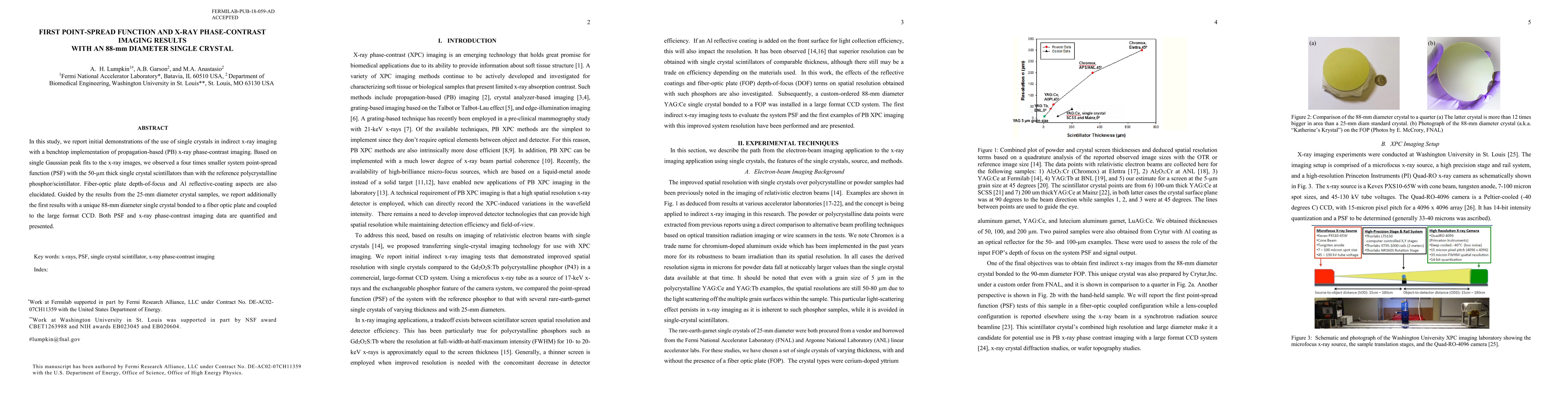
In this study, we report initial demonstrations of the use of single crystals in indirect x-ray imaging with a benchtop implementation of propagation-based (PB) x-ray phase contrast imaging. Based on single Gaussian peak fits to the x-ray images, we observed a four times smaller system point-spread function (PSF) with the 50-{\mu}m thick single crystal scintillators than with the reference polycrystalline phosphor/scintillator. Fiber-optic plate depth-of-focus and Al reflective-coating aspects are also elucidated. Guided by the results from the 25-mm diameter crystal samples, we report additionally the first results with a unique 88-mm diameter single crystal bonded to a fiber optic plate and coupled to the large format CCD. Both PSF and x-ray phase contrast imaging data are quantified and presented.
AI Key Findings
Get AI-generated insights about this paper's methodology, results, significance, and more — seven facets brought into focus.
Impact
Paper Details
PDF Preview
Key Terms
Citation Network
Current paper (gray), citations (green), references (blue)
Display is limited for performance on very large graphs.
Discussion 0