Fluorescence microscopy of single autofluorescent proteins for cellular biology
Publication
Metrics
AI Quick Summary
This paper reviews the use of autofluorescent proteins like GFP and DsRed mutants for single-molecule imaging in cellular biology, comparing their photophysical properties. It also explores the potential of two-photon excitation and Fluorescence Correlation Spectroscopy for studying individual autofluorescent proteins in live cells, highlighting the first use of the more photostable citrine variant fused to a receptor protein.
Paper Preview
Abstract
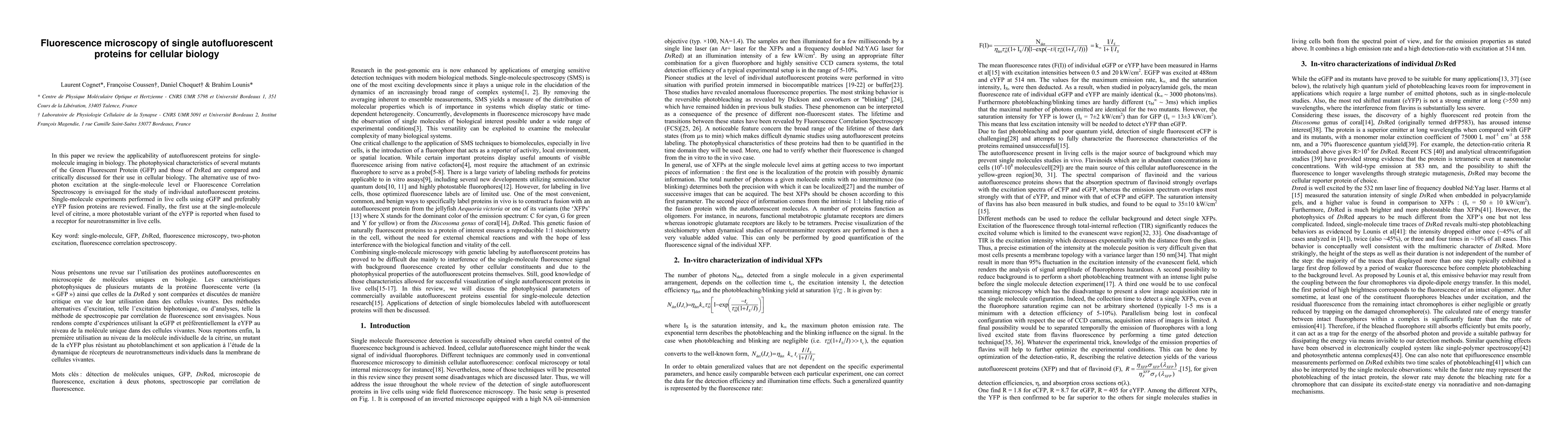
In this paper we review the applicability of autofluorescent proteins for single-molecule imaging in biology. The photophysical characteristics of several mutants of the Green Fluorescent Protein (GFP) and those of DsRed are compared and critically discussed for their use in cellular biology. The alternative use of two-photon excitation at the single-molecule level or Fluorescence Correlation Spectroscopy is envisaged for the study of individual autofluorescent proteins. Single-molecule experiments performed in live cells using eGFP and preferably eYFP fusion proteins are reviewed. Finally, the first use at the single-molecule level of citrine, a more photostable variant of the eYFP is reported when fused to a receptor for neurotransmitter in live cells.
AI Key Findings
Get AI-generated insights about this paper's methodology, results, significance, and more — seven facets brought into focus.
Impact
Paper Details
PDF Preview
Key Terms
Citation Network
Current paper (gray), citations (green), references (blue)
Display is limited for performance on very large graphs.
Discussion 0