ArxivLens

Publication

Published: Jan 19, 2026
Categories: cs.CV, cs.AI, cs.LG

Metrics

Source: ArXiv

Quick Answers

What methodology did the authors use?

The research employed a U-Net CNN architecture combined with masked autoencoders (MAE) and strategies leveraging anatomical priors and neuroimaging domain knowledge. The models were trained on large-scale 3D brain MRI datasets from the SSL3D and FOMO25 challenges, with evaluations across segmentation, classification, and regression tasks. More in Methodology →

What are the key results?

The proposed CNN-based models outperformed transformer-based approaches in both segmentation and classification tasks, achieving 2.5% higher average Dice and 8% higher accuracy. — Models trained 1-2 orders of magnitude faster and were 10× smaller than competing transformer-based approaches. More in Key Results →

Why is this work significant?

This research advances medical image analysis by demonstrating the effectiveness of CNN-based foundation models in overcoming challenges like data sparsity and protocol variability. It provides a practical framework for developing efficient, domain-aware models for brain MRI analysis, which is critical for clinical applications. More in Significance →

What are the main limitations?

The results are specific to brain MRI and may not generalize to other medical imaging modalities. — The study focuses on pre-training and fine-tuning strategies but does not address long-term model maintenance or updates. More in Limitations →

Paper Preview

Abstract

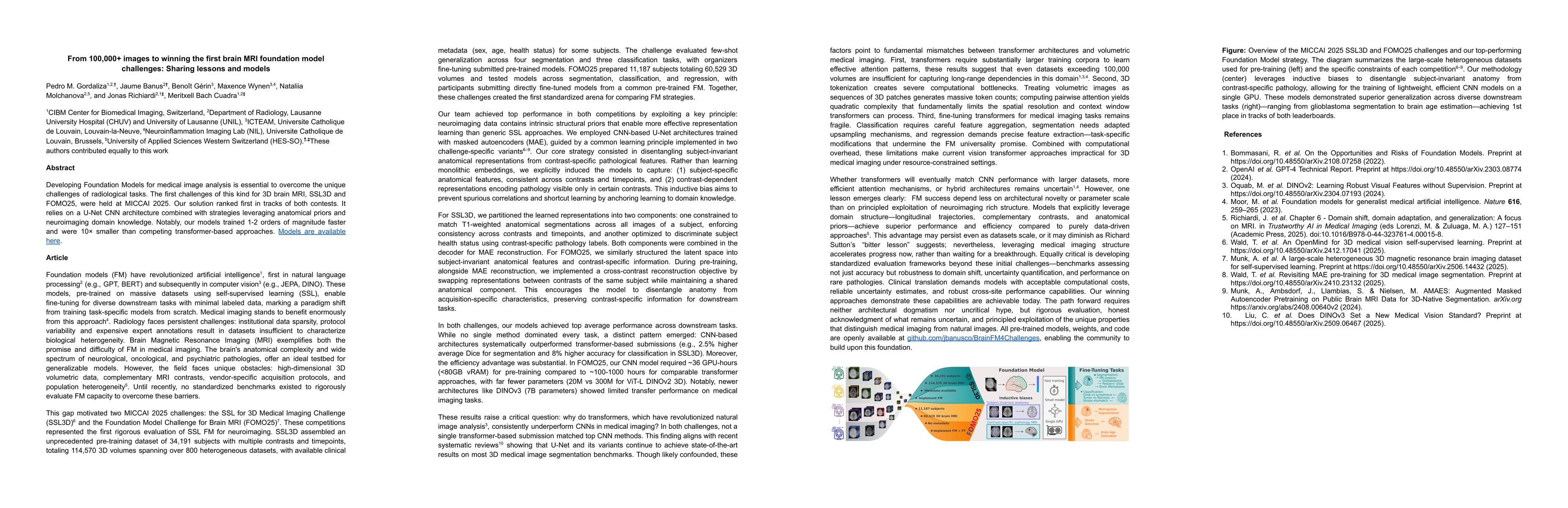
Developing Foundation Models for medical image analysis is essential to overcome the unique challenges of radiological tasks. The first challenges of this kind for 3D brain MRI, SSL3D and FOMO25, were held at MICCAI 2025. Our solution ranked first in tracks of both contests. It relies on a U-Net CNN architecture combined with strategies leveraging anatomical priors and neuroimaging domain knowledge. Notably, our models trained 1-2 orders of magnitude faster and were 10 times smaller than competing transformer-based approaches. Models are available here: https://github.com/jbanusco/BrainFM4Challenges.

Key Findings, in focus

Seven facets of this paper, analysed and brought into focus by AI.

AI-generated · verify against source Generated Jan 23, 2026
In focus · one-line synthesis

This research advances medical image analysis by demonstrating the effectiveness of CNN-based foundation models in overcoming challenges like data sparsity and protocol variability.

01
MethodologyHow they did it

The research employed a U-Net CNN architecture combined with masked autoencoders (MAE) and strategies leveraging anatomical priors and neuroimaging domain knowledge. The models were trained on large-scale 3D brain MRI datasets from the SSL3D and FOMO25 challenges, with evaluations across segmentation, classification, and regression tasks.

02
ResultsWhat they found
  • The proposed CNN-based models outperformed transformer-based approaches in both segmentation and classification tasks, achieving 2.5% higher average Dice and 8% higher accuracy.
  • Models trained 1-2 orders of magnitude faster and were 10× smaller than competing transformer-based approaches.
  • The approach demonstrated superior generalization across diverse downstream tasks, achieving top performance in both SSL3D and FOMO25 challenges.
03
SignificanceWhy it matters

This research advances medical image analysis by demonstrating the effectiveness of CNN-based foundation models in overcoming challenges like data sparsity and protocol variability. It provides a practical framework for developing efficient, domain-aware models for brain MRI analysis, which is critical for clinical applications.

04
Technical ContributionsWhat's new technically

The technical contribution lies in the integration of anatomical priors and neuroimaging domain knowledge into CNN-based foundation models, enabling more effective representation learning and efficient model training.

05
NoveltyWhat sets it apart

The novelty is in the explicit disentanglement of subject-invariant anatomical features from contrast-specific pathological features, combined with the use of masked autoencoders guided by domain-specific learning principles.

06
LimitationsWhat to be cautious about
  • The results are specific to brain MRI and may not generalize to other medical imaging modalities.
  • The study focuses on pre-training and fine-tuning strategies but does not address long-term model maintenance or updates.
07
Future WorkWhere it goes next
  • Exploring hybrid architectures that combine CNNs and transformers for improved performance.
  • Developing standardized evaluation frameworks that assess robustness to domain shifts and uncertainty quantification.
  • Investigating the impact of larger datasets and more efficient attention mechanisms on transformer performance in medical imaging.

Paper Details

Paper ID: 2601.13166
Comments: Work presented at the SSL3D Challenge (1st place, ResEnc-L track) and FOMO Challenge (1st place, Methods track) on Brain MRI Foundation Models at MICCAI 2025
Categories:
cs.CV cs.AI cs.LG

How to Cite This Paper

Reference manager: RIS EndNote


Discussion 0

This website uses cookies to ensure you get the best experience. Learn more