Academic Profile
Statistics
Similar Authors
Papers on arXiv

Uncertainty quantification (UQ) has become critical for evaluating the reliability of artificial intelligence systems, especially in medical image segmentation. This study addresses the interpretabili...
In recent years, explainable methods for artificial intelligence (XAI) have tried to reveal and describe models' decision mechanisms in the case of classification tasks. However, XAI for semantic segm...
The thalamus is a subcortical structure of central importance to brain function, which is organized in smaller nuclei with specialized roles. Despite significant functional and clinical relevance, l...
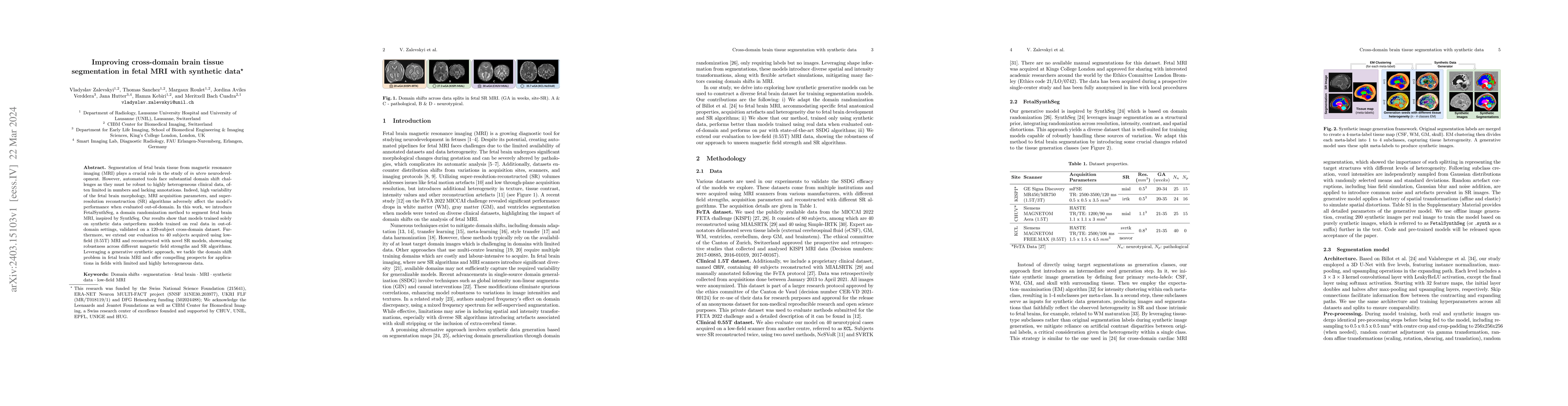
Segmentation of fetal brain tissue from magnetic resonance imaging (MRI) plays a crucial role in the study of in utero neurodevelopment. However, automated tools face substantial domain shift challe...
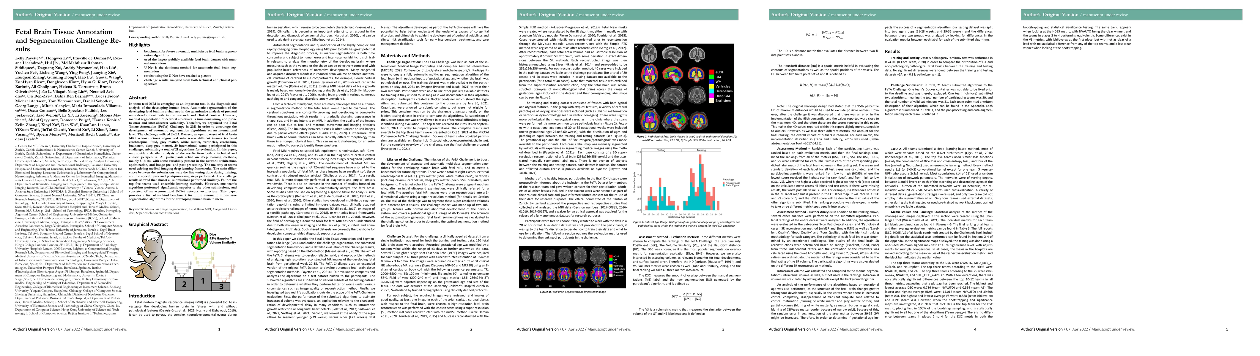
Segmentation is a critical step in analyzing the developing human fetal brain. There have been vast improvements in automatic segmentation methods in the past several years, and the Fetal Brain Tiss...
Deep learning models have shown great promise in estimating tissue microstructure from limited diffusion magnetic resonance imaging data. However, these models face domain shift challenges when test...
This paper explores uncertainty quantification (UQ) as an indicator of the trustworthiness of automated deep-learning (DL) tools in the context of white matter lesion (WML) segmentation from magneti...
The brain white matter consists of a set of tracts that connect distinct regions of the brain. Segmentation of these tracts is often needed for clinical and research studies. Diffusion-weighted MRI ...
Accurate segmentation of thalamic nuclei, crucial for understanding their role in healthy cognition and in pathologies, is challenging to achieve on standard T1-weighted (T1w) magnetic resonance ima...
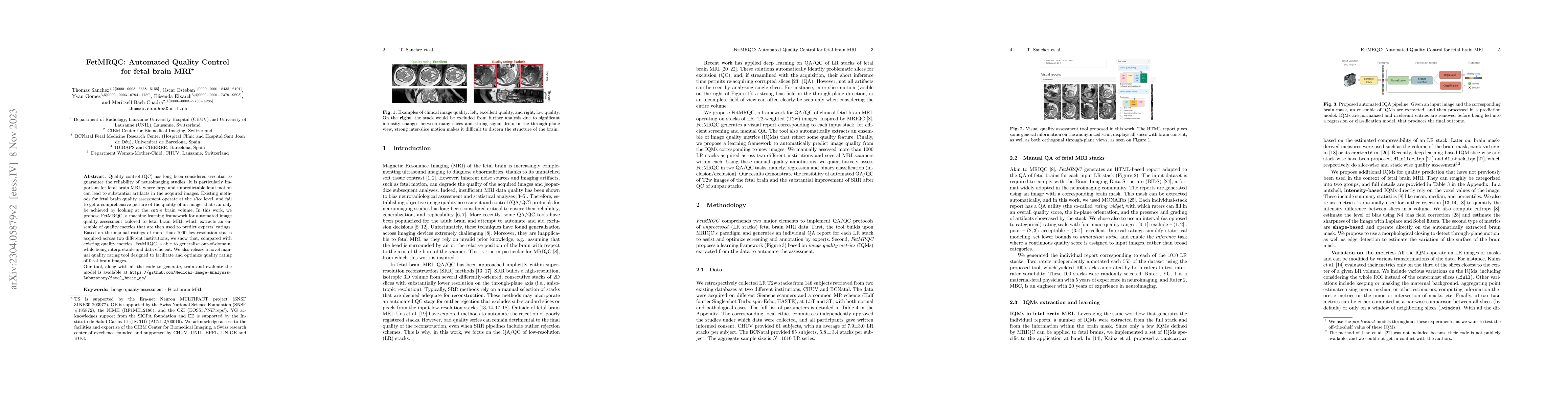
Quality control (QC) has long been considered essential to guarantee the reliability of neuroimaging studies. It is particularly important for fetal brain MRI, where large and unpredictable fetal mo...
The development of automatic segmentation techniques for medical imaging tasks requires assessment metrics to fairly judge and rank such approaches on benchmarks. The Dice Similarity Coefficient (DS...
The benefits, opportunities and growing availability of ultra-high field magnetic resonance imaging (MRI) for humans have prompted an expansion in research and development efforts towards increasing...
Quantitative analysis of in utero human brain development is crucial for abnormal characterization. Magnetic resonance image (MRI) segmentation is therefore an asset for quantitative analysis. Howev...
Tuning the regularization hyperparameter $\alpha$ in inverse problems has been a longstanding problem. This is particularly true in the case of fetal brain magnetic resonance imaging, where an isotr...
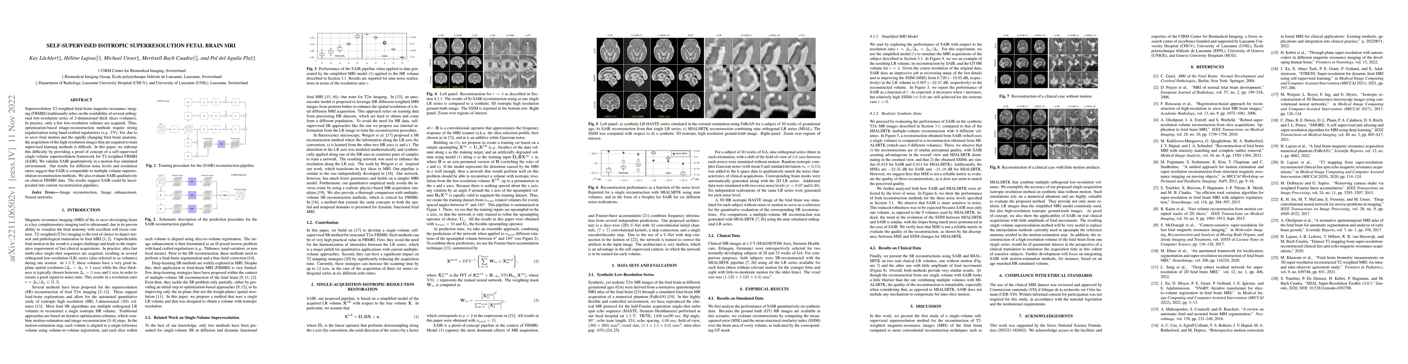
Superresolution T2-weighted fetal-brain magnetic-resonance imaging (FBMRI) traditionally relies on the availability of several orthogonal low-resolution series of 2-dimensional thick slices (volumes...
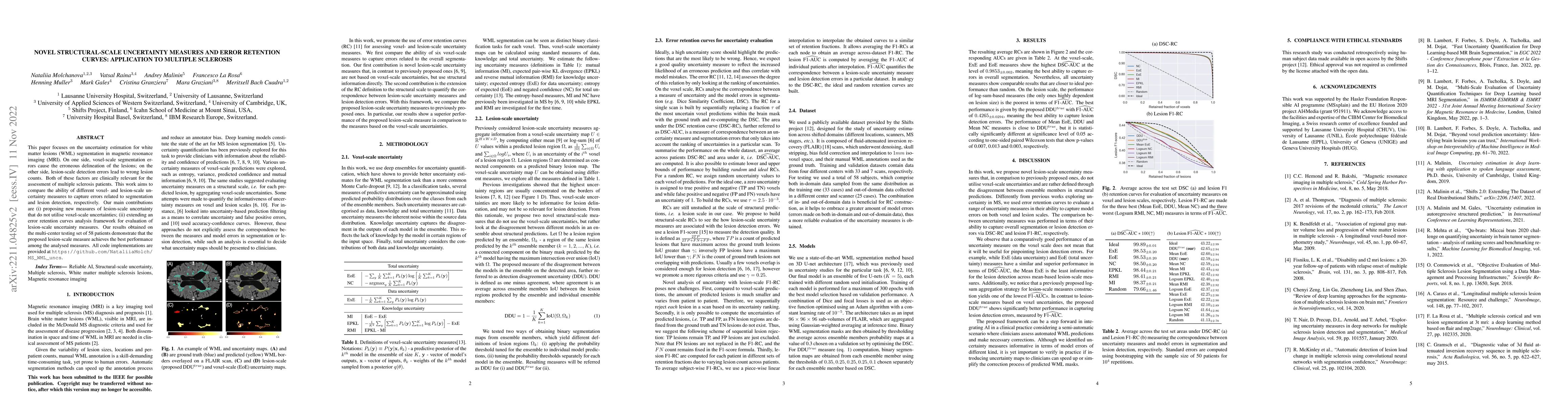
This paper focuses on the uncertainty estimation for white matter lesions (WML) segmentation in magnetic resonance imaging (MRI). On one side, voxel-scale segmentation errors cause the erroneous del...
Gliomas are the most frequent primary brain tumors in adults. Glioma change detection aims at finding the relevant parts of the image that change over time. Although Deep Learning (DL) shows promisi...
Resting-state functional Magnetic Resonance Imaging (fMRI) is a powerful imaging technique for studying functional development of the brain in utero. However, unpredictable and excessive movement of...
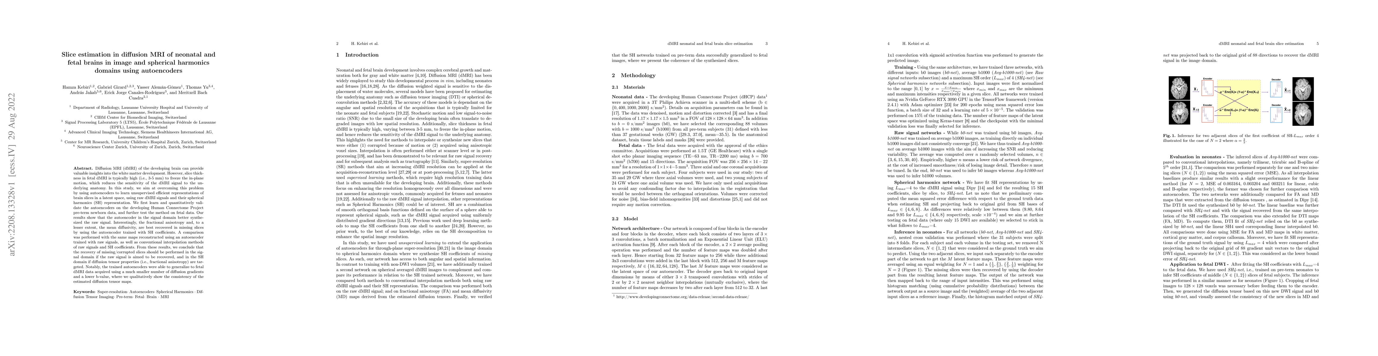
Diffusion MRI (dMRI) of the developing brain can provide valuable insights into the white matter development. However, slice thickness in fetal dMRI is typically high (i.e., 3-5 mm) to freeze the in...
The fetal cortical plate (CP) undergoes drastic morphological changes during the in utero development. Therefore, CP growth and folding patterns are key indicator in the assessment of the brain deve...
Distributional shift, or the mismatch between training and deployment data, is a significant obstacle to the usage of machine learning in high-stakes industrial applications, such as autonomous driv...
In-utero fetal MRI is emerging as an important tool in the diagnosis and analysis of the developing human brain. Automatic segmentation of the developing fetal brain is a vital step in the quantitat...
Deep learning methods have become the state of the art for undersampled MR reconstruction. Particularly for cases where it is infeasible or impossible for ground truth, fully sampled data to be acqu...
The current multiple sclerosis (MS) diagnostic criteria lack specificity, and this may lead to misdiagnosis, which remains an issue in present-day clinical practice. In addition, conventional biomar...
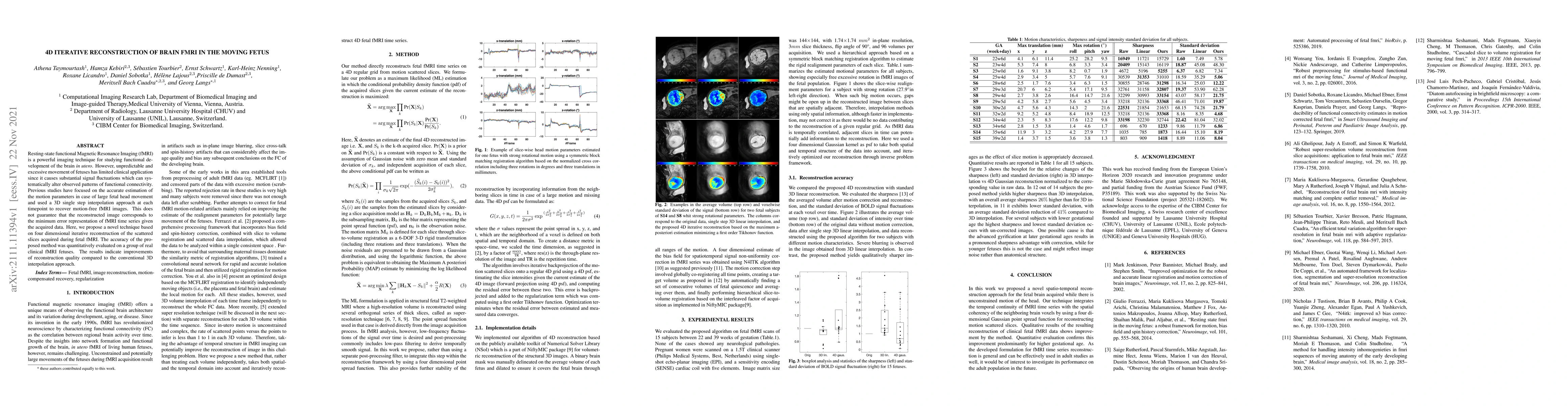
Resting-state functional Magnetic Resonance Imaging (fMRI) is a powerful imaging technique for studying functional development of the brain in utero. However, unpredictable and excessive movement of...
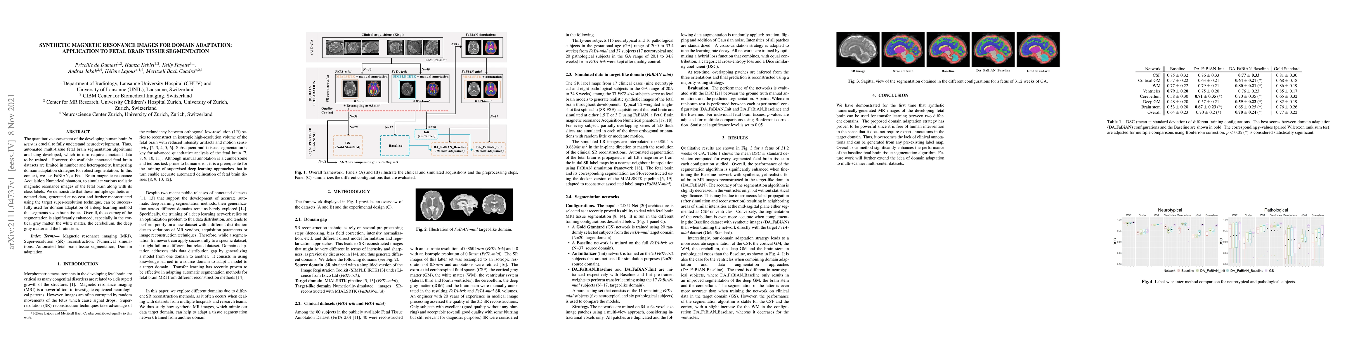
The quantitative assessment of the developing human brain in utero is crucial to fully understand neurodevelopment. Thus, automated multi-tissue fetal brain segmentation algorithms are being develop...
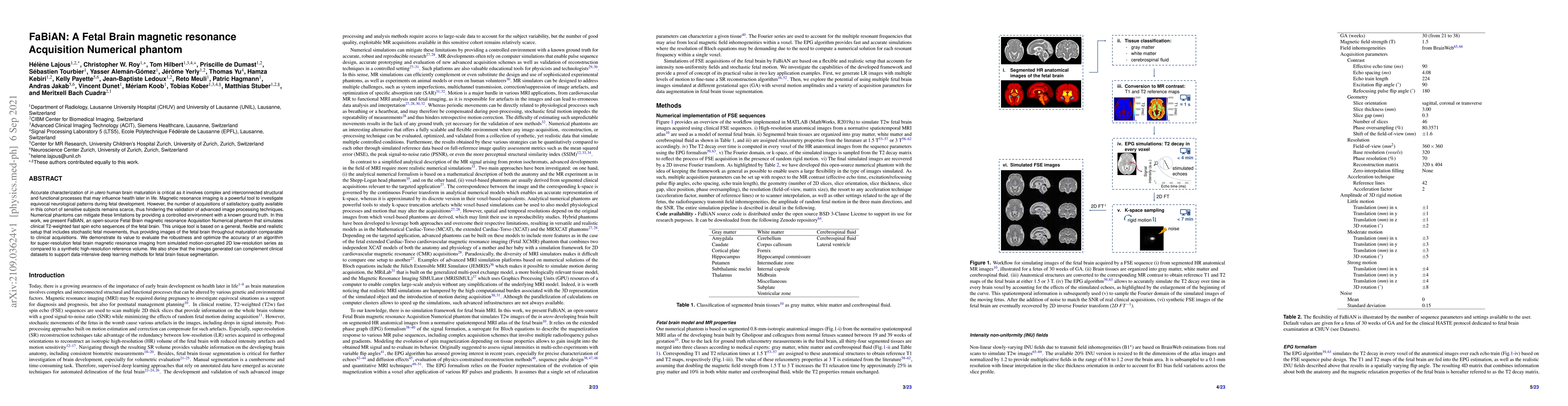
Accurate characterization of in utero human brain maturation is critical as it involves complex and interconnected structural and functional processes that may influence health later in life. Magnet...
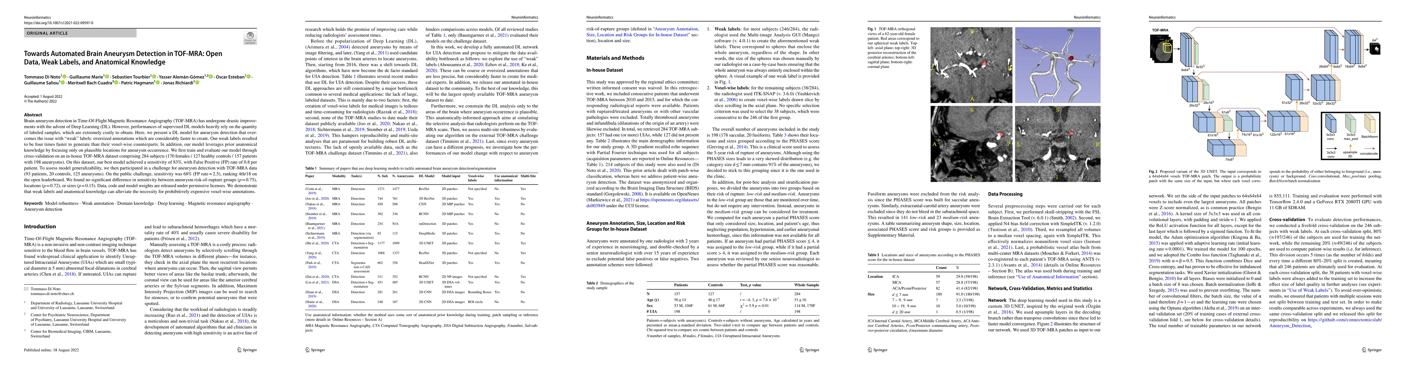
Brain aneurysm detection in Time-Of-Flight Magnetic Resonance Angiography (TOF-MRA) has undergone drastic improvements with the advent of Deep Learning (DL). However, performances of supervised DL m...
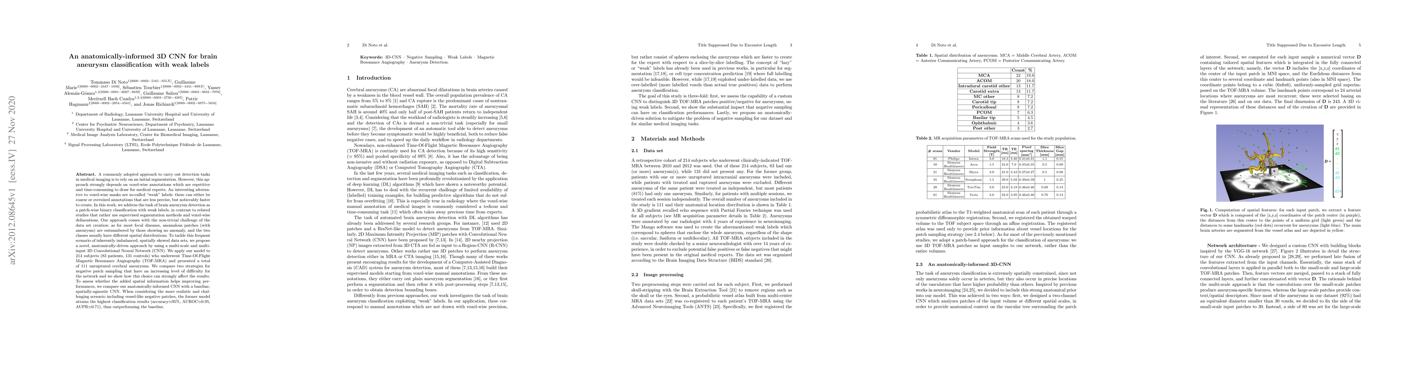
A commonly adopted approach to carry out detection tasks in medical imaging is to rely on an initial segmentation. However, this approach strongly depends on voxel-wise annotations which are repetit...
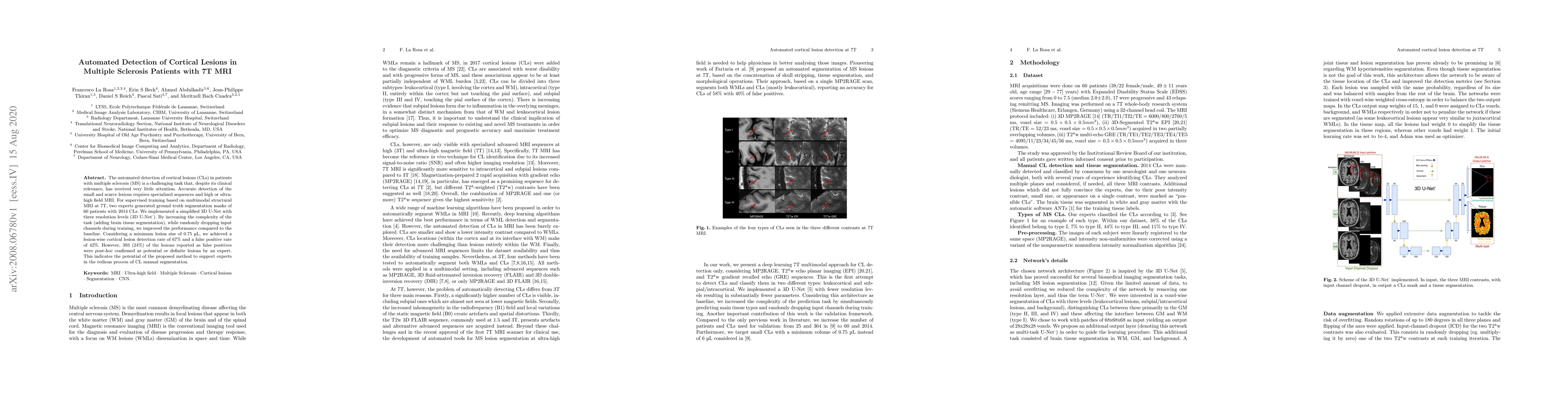
The automated detection of cortical lesions (CLs) in patients with multiple sclerosis (MS) is a challenging task that, despite its clinical relevance, has received very little attention. Accurate de...
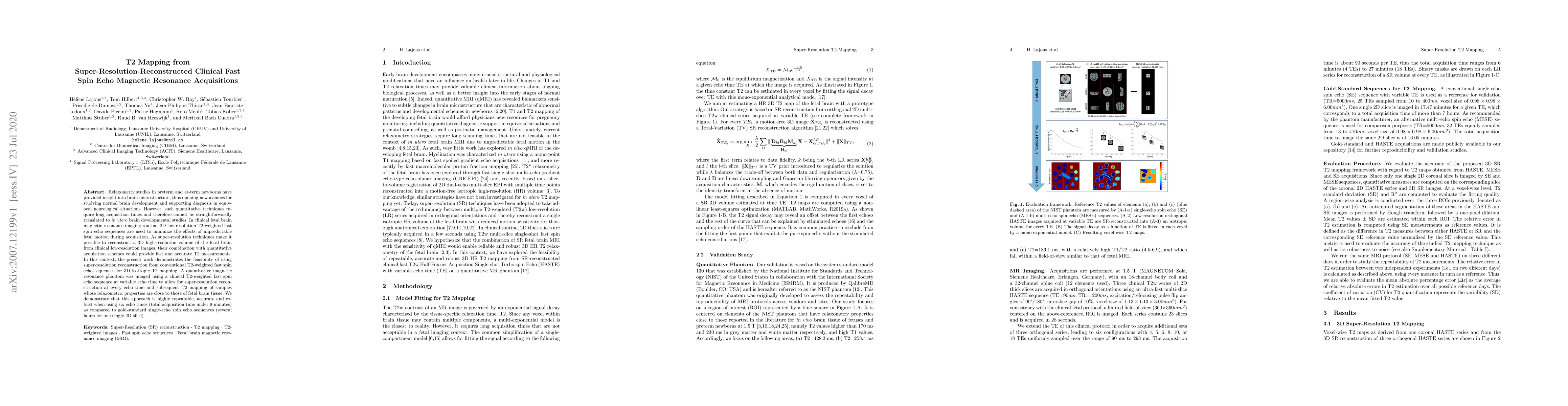
Relaxometry studies in preterm and at-term newborns have provided insight into brain microstructure, thus opening new avenues for studying normal brain development and supporting diagnosis in equivo...
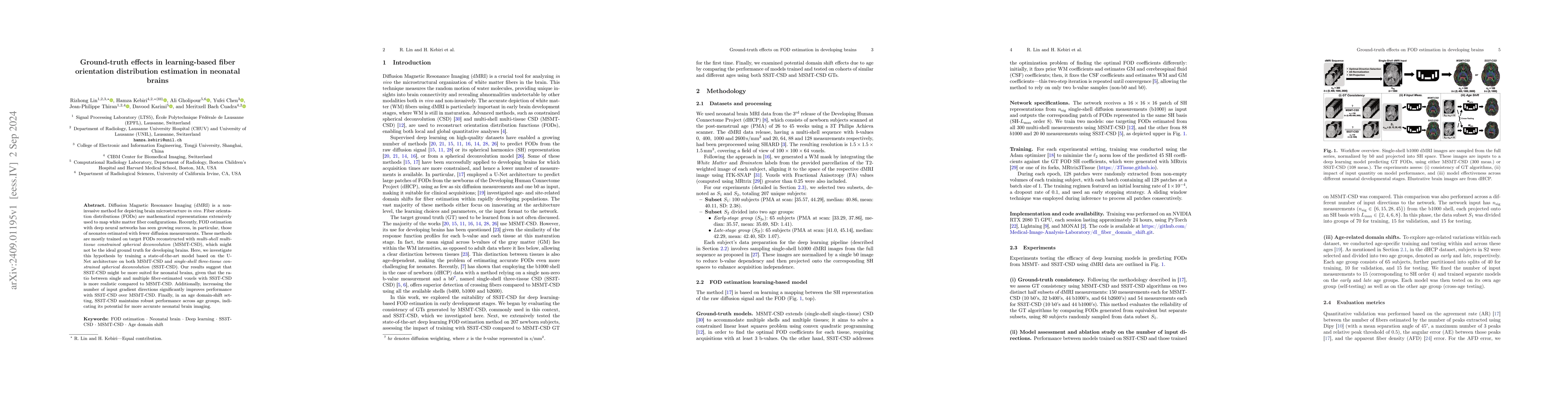
Diffusion Magnetic Resonance Imaging (dMRI) is a non-invasive method for depicting brain microstructure in vivo. Fiber orientation distributions (FODs) are mathematical representations extensively use...
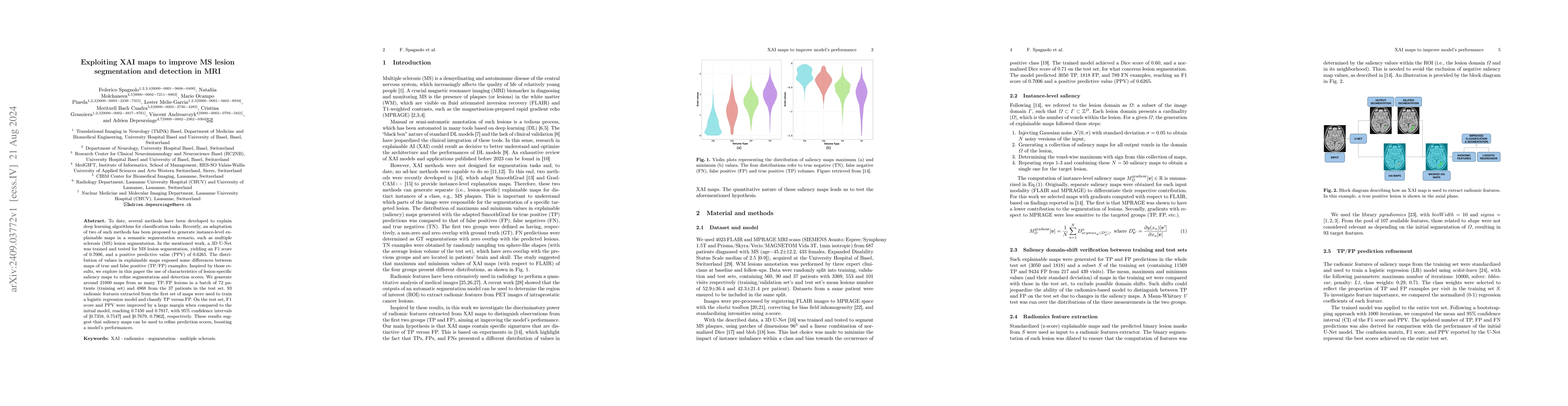
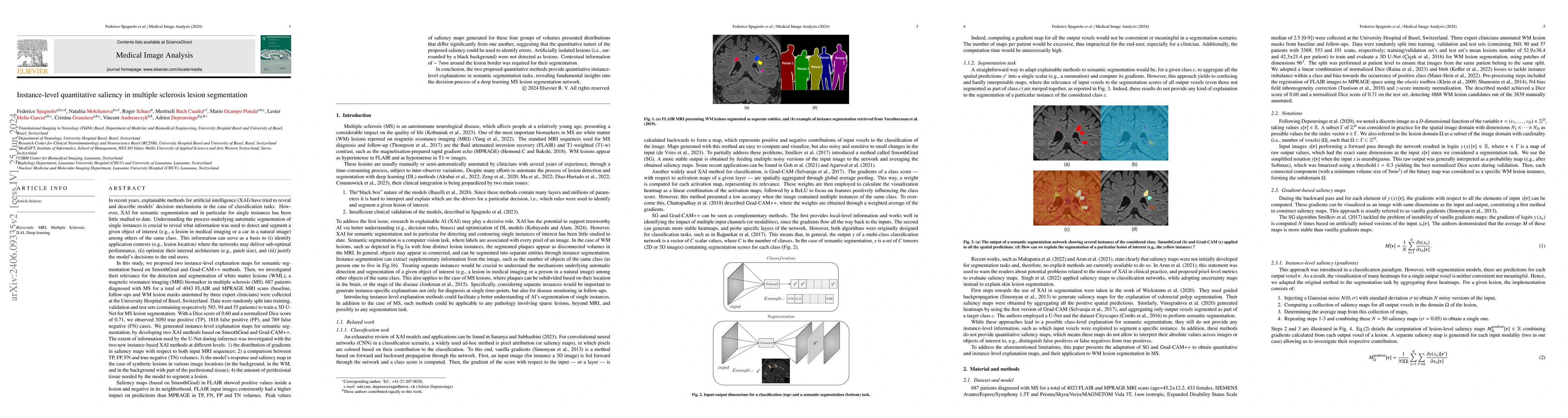
To date, several methods have been developed to explain deep learning algorithms for classification tasks. Recently, an adaptation of two of such methods has been proposed to generate instance-level e...
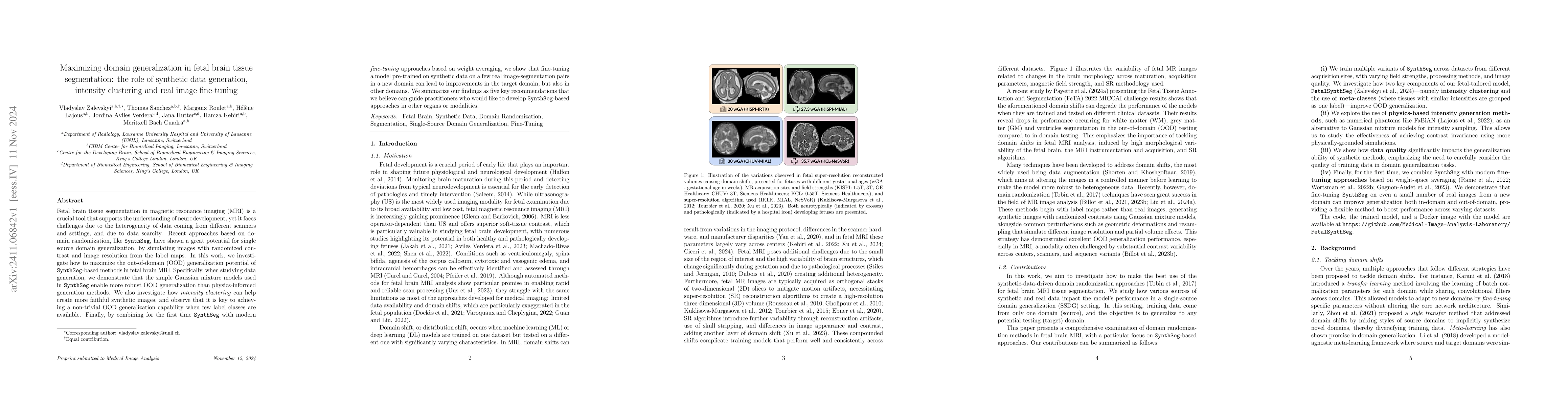
Fetal brain tissue segmentation in magnetic resonance imaging (MRI) is a crucial tool that supports the understanding of neurodevelopment, yet it faces challenges due to the heterogeneity of data comi...
Despite the plethora of AI-based algorithms developed for anomaly detection in radiology, subsequent integration into clinical setting is rarely evaluated. In this work, we assess the applicability an...
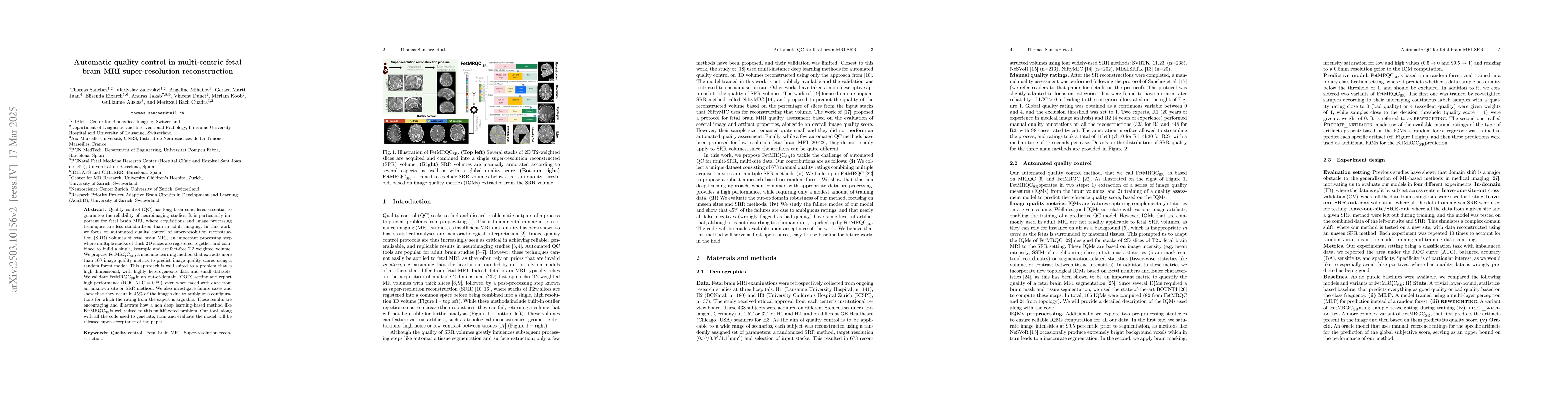
Quality control (QC) has long been considered essential to guarantee the reliability of neuroimaging studies. It is particularly important for fetal brain MRI, where acquisitions and image processing ...
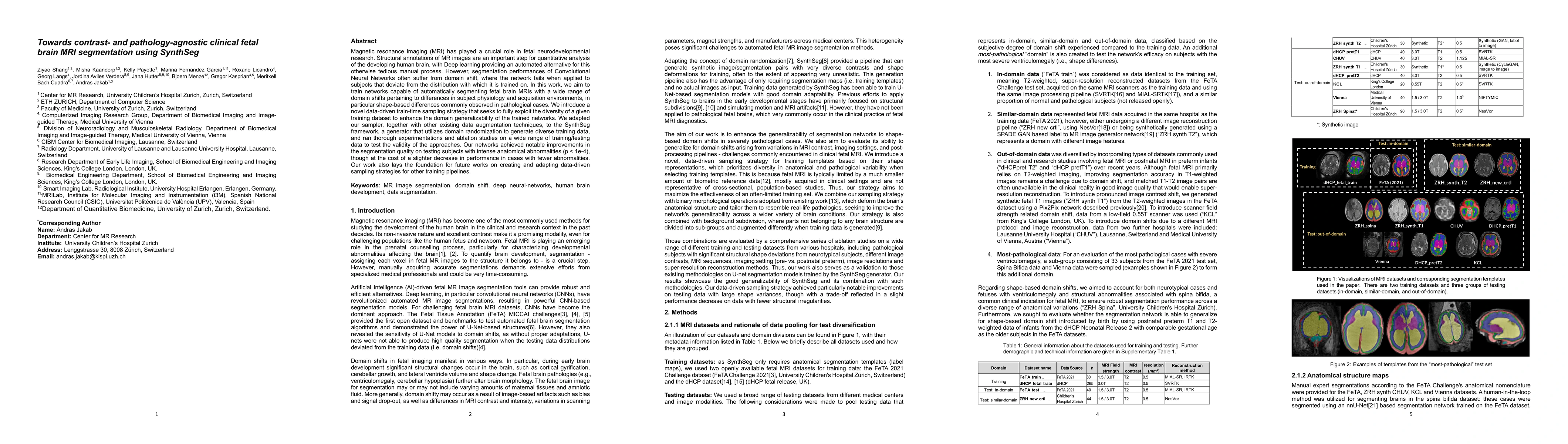
Magnetic resonance imaging (MRI) has played a crucial role in fetal neurodevelopmental research. Structural annotations of MR images are an important step for quantitative analysis of the developing h...
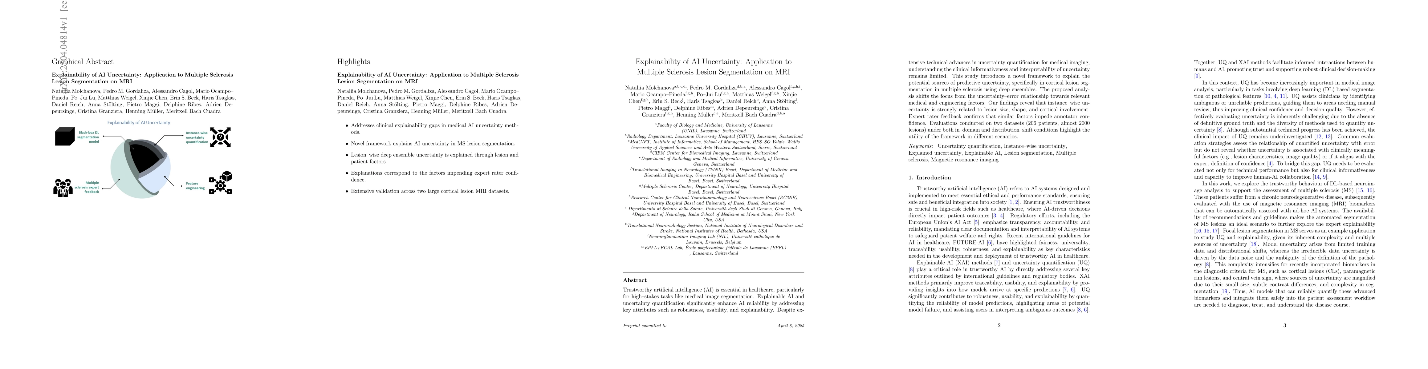
Trustworthy artificial intelligence (AI) is essential in healthcare, particularly for high-stakes tasks like medical image segmentation. Explainable AI and uncertainty quantification significantly enh...
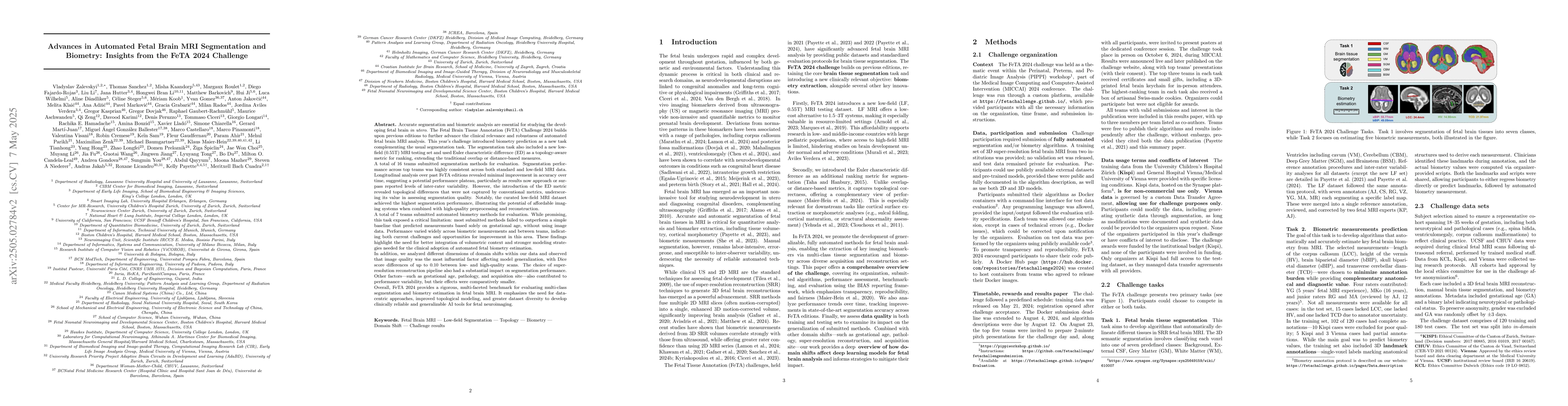
Accurate fetal brain tissue segmentation and biometric analysis are essential for studying brain development in utero. The FeTA Challenge 2024 advanced automated fetal brain MRI analysis by introducin...
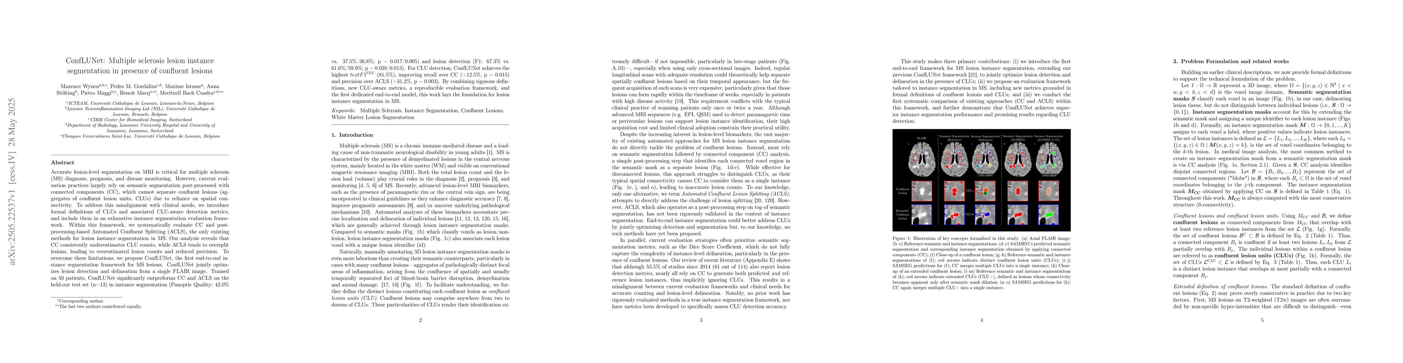
Accurate lesion-level segmentation on MRI is critical for multiple sclerosis (MS) diagnosis, prognosis, and disease monitoring. However, current evaluation practices largely rely on semantic segmentat...
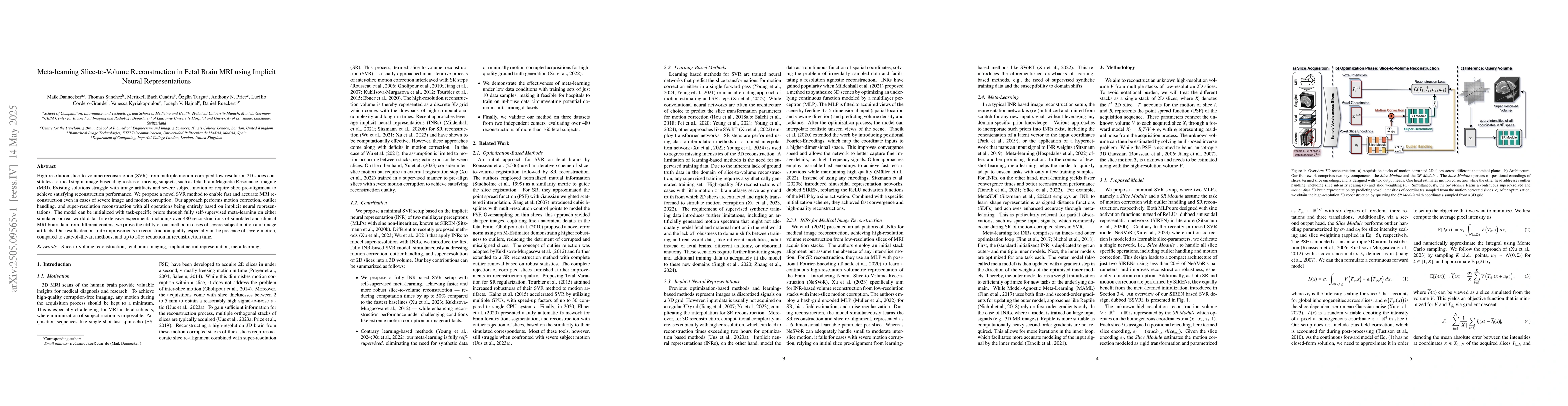
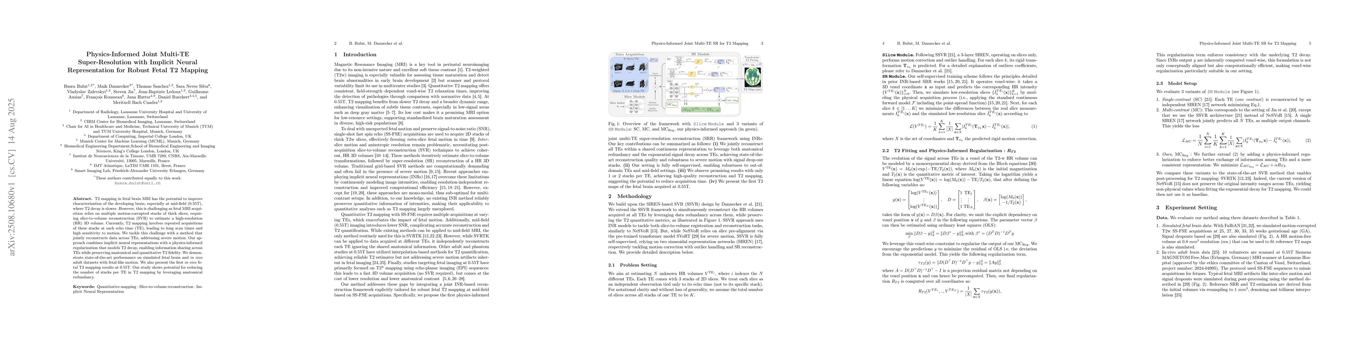
High-resolution slice-to-volume reconstruction (SVR) from multiple motion-corrupted low-resolution 2D slices constitutes a critical step in image-based diagnostics of moving subjects, such as fetal br...
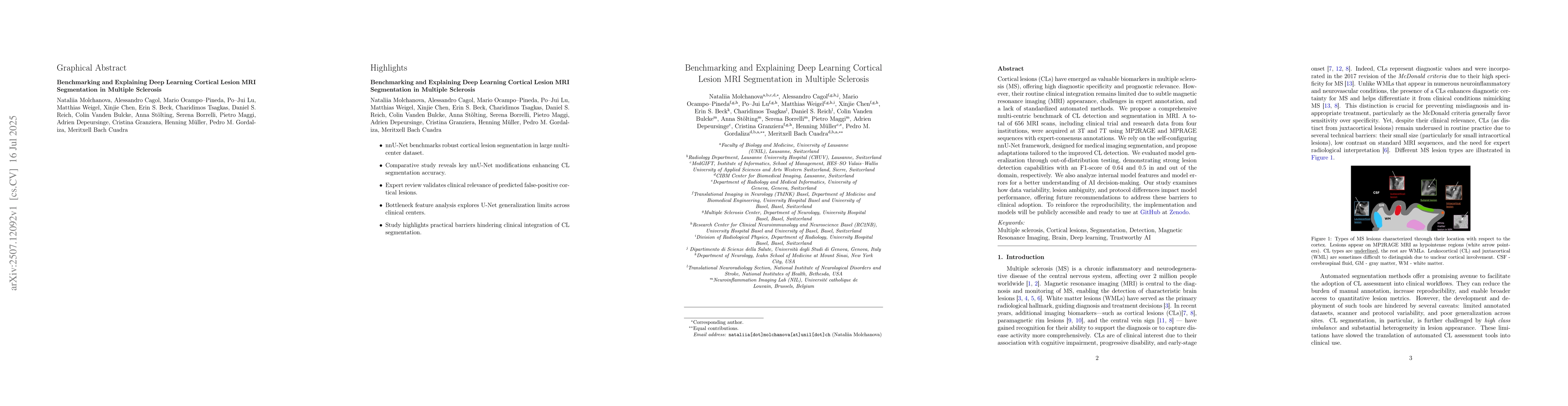
Cortical lesions (CLs) have emerged as valuable biomarkers in multiple sclerosis (MS), offering high diagnostic specificity and prognostic relevance. However, their routine clinical integration remain...
T2 mapping in fetal brain MRI has the potential to improve characterization of the developing brain, especially at mid-field (0.55T), where T2 decay is slower. However, this is challenging as fetal MR...
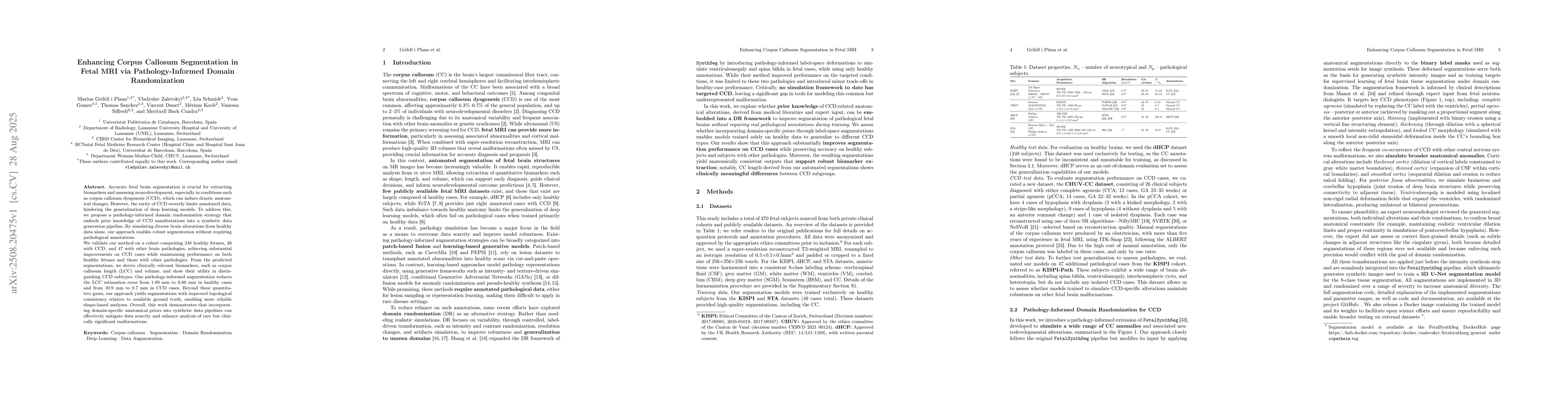
Accurate fetal brain segmentation is crucial for extracting biomarkers and assessing neurodevelopment, especially in conditions such as corpus callosum dysgenesis (CCD), which can induce drastic anato...
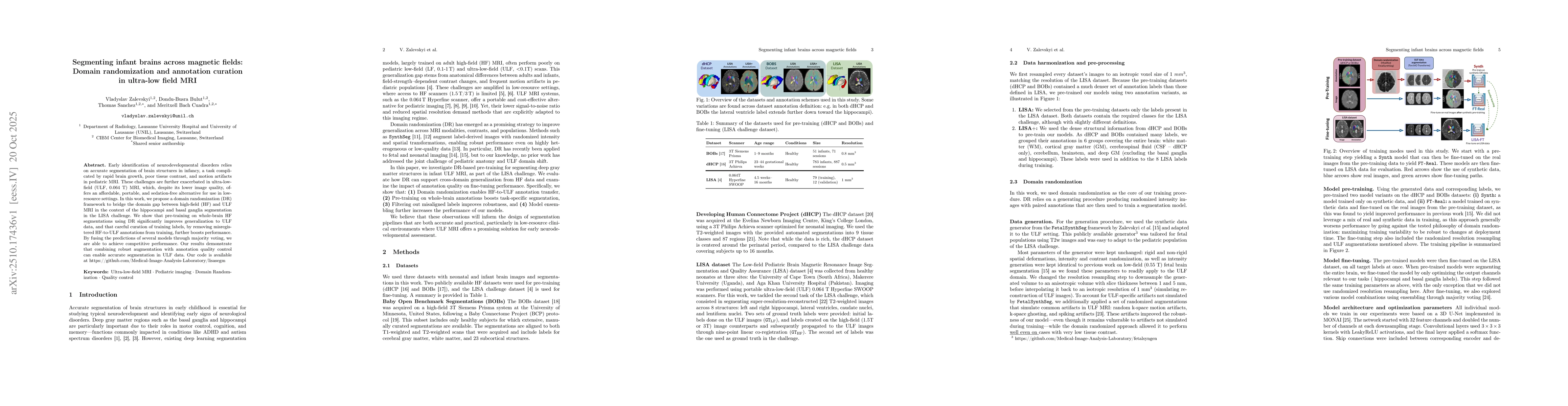
Early identification of neurodevelopmental disorders relies on accurate segmentation of brain structures in infancy, a task complicated by rapid brain growth, poor tissue contrast, and motion artifact...
Low-field T2 mapping MRI can democratize neuropediatric imaging by improving accessibility and providing quantitative biomarkers of brain development. \textbf{Purpose:} To evaluate the feasibility of ...
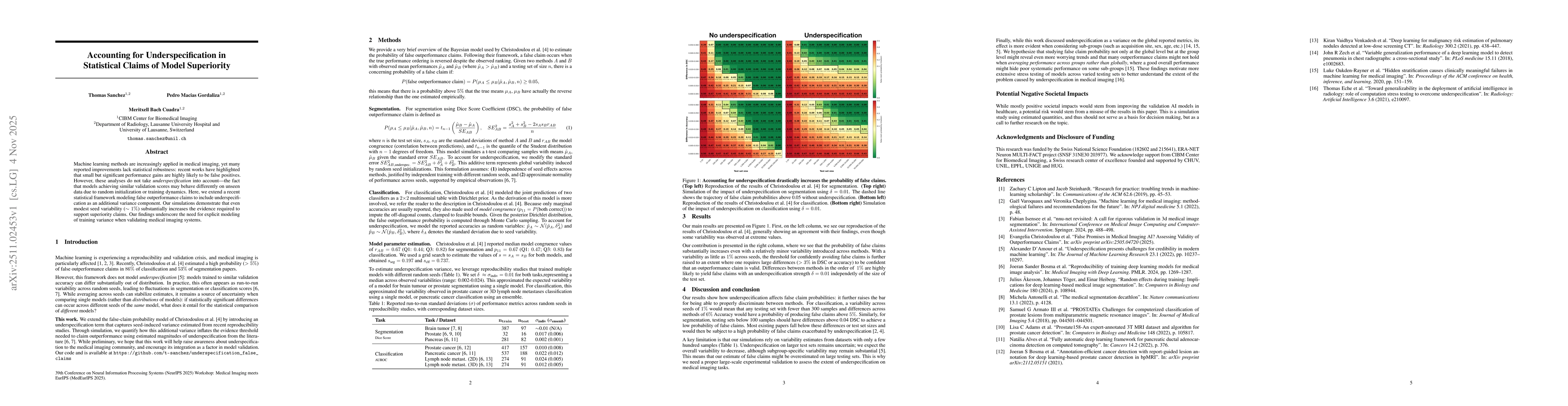
Machine learning methods are increasingly applied in medical imaging, yet many reported improvements lack statistical robustness: recent works have highlighted that small but significant performance g...
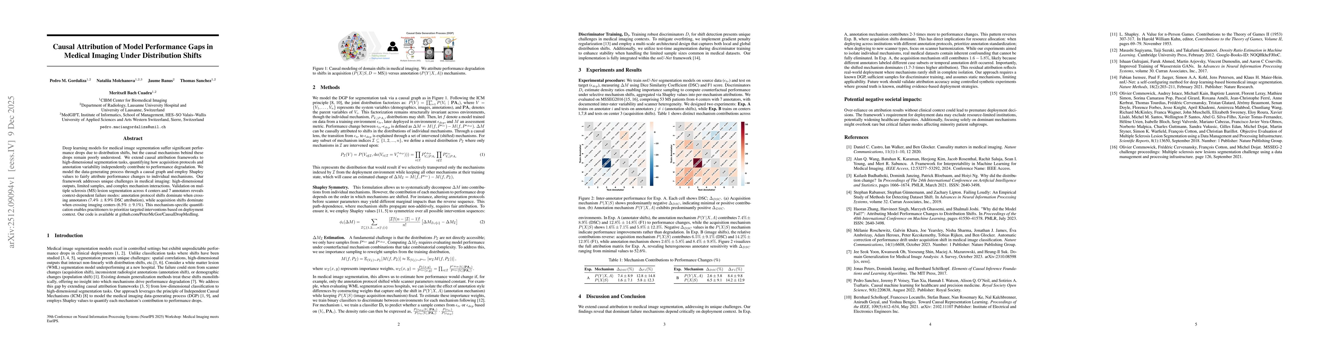
Deep learning models for medical image segmentation suffer significant performance drops due to distribution shifts, but the causal mechanisms behind these drops remain poorly understood. We extend ca...
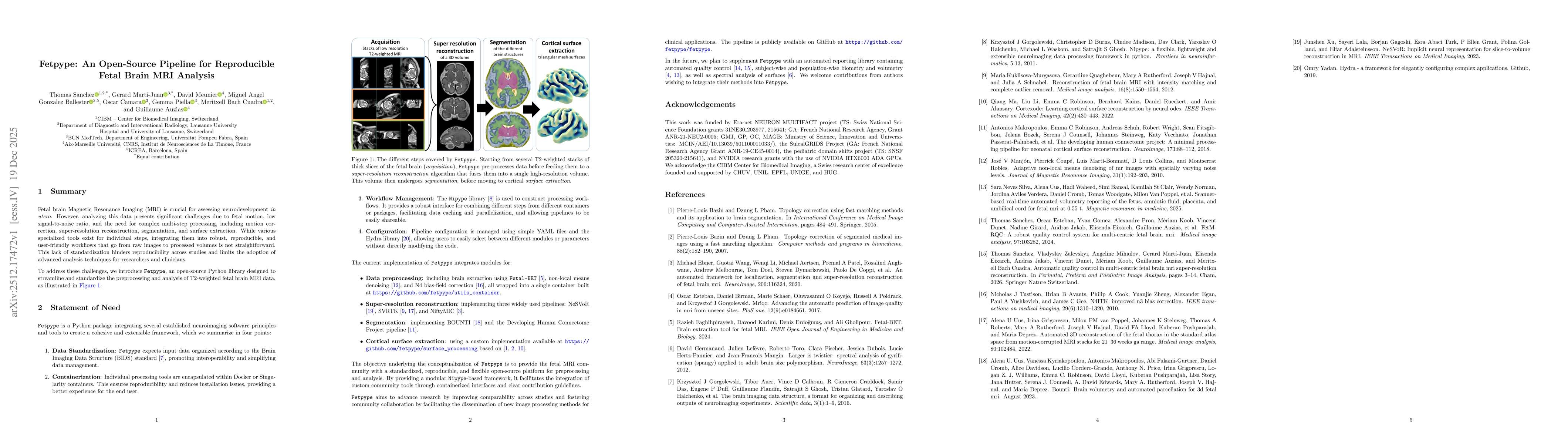
Fetal brain Magnetic Resonance Imaging (MRI) is crucial for assessing neurodevelopment in utero. However, analyzing this data presents significant challenges due to fetal motion, low signal-to-noise r...
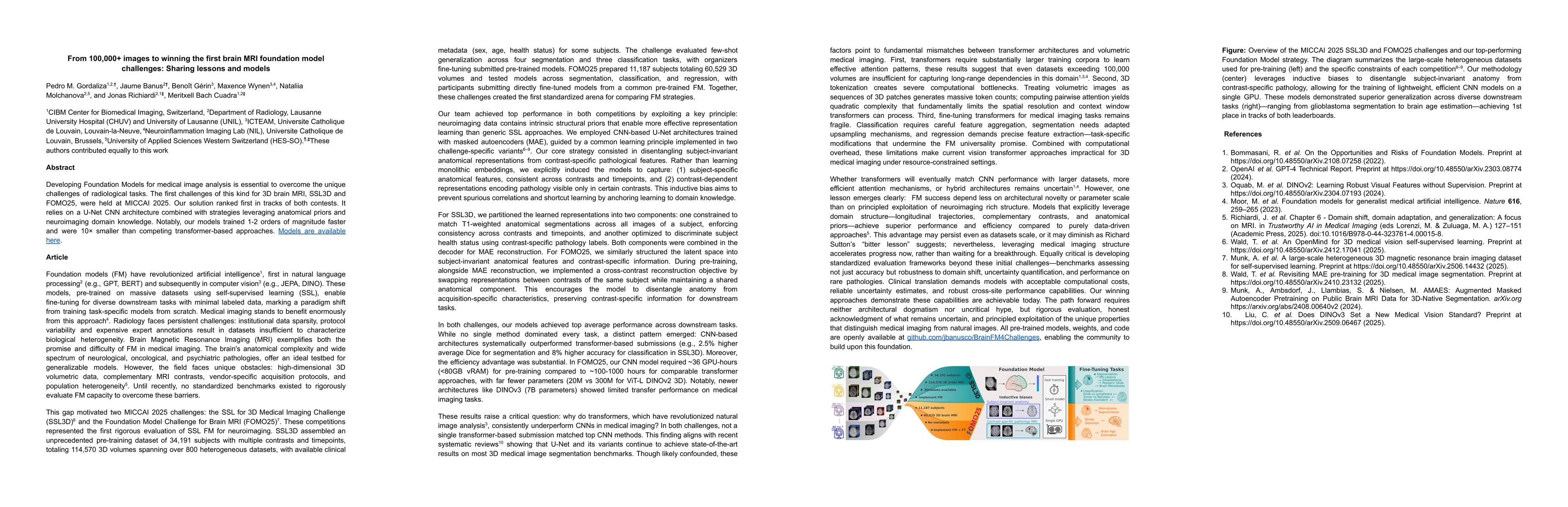
Developing Foundation Models for medical image analysis is essential to overcome the unique challenges of radiological tasks. The first challenges of this kind for 3D brain MRI, SSL3D and FOMO25, were...
Clinical deployment of automated brain MRI analysis faces a fundamental challenge: clinical data is heterogeneous and noisy, and high-quality labels are prohibitively costly to obtain. Self-supervised...