This paper evaluates various 7T MRI techniques for distinguishing human thalamic nuclei, finding that quantitative susceptibility mapping (QSM) and optimized T$_{1}$-weighted MP2RAGE provide the most detailed differentiation of thalamic nuclei, offering unique and complementary insights for improved imaging and automatic segmentation.
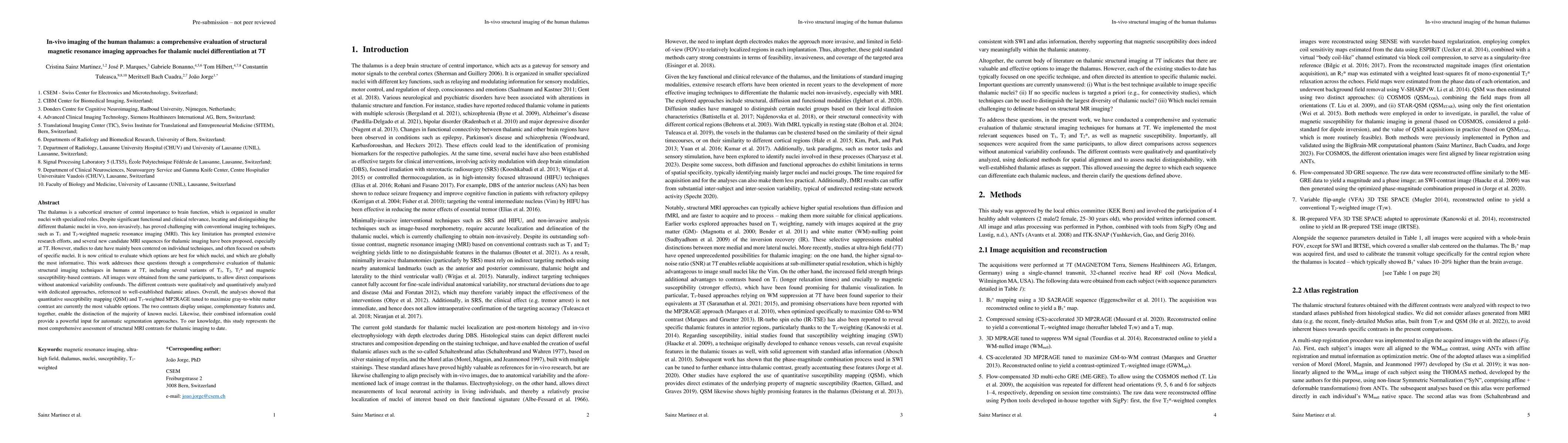
The thalamus is a subcortical structure of central importance to brain function, which is organized in smaller nuclei with specialized roles. Despite significant functional and clinical relevance, locating and distinguishing the different thalamic nuclei in vivo, non-invasively, has proved challenging with conventional imaging techniques, such as T$_{1}$ and T$_{2}$-weighted magnetic resonance imaging (MRI). This key limitation has prompted extensive research efforts, and several new candidate MRI sequences for thalamic imaging have been proposed, especially at 7T. However, studies to date have mainly been centered on individual techniques, and often focused on subsets of specific nuclei. It is now critical to evaluate which options are best for which nuclei, and which are globally the most informative. This work addresses these questions through a comprehensive evaluation of thalamic structural imaging techniques in humans at 7T, including several variants of T$_{1}$, T$_{2}$, T$_{2}$* and magnetic susceptibility-based contrasts. All images were obtained from the same participants, to allow direct comparisons without anatomical variability confounds. The different contrasts were qualitatively and quantitatively analyzed with dedicated approaches, referenced to well-established thalamic atlases. Overall, the analyses showed that quantitative susceptibility mapping (QSM) and T$_{1}$-weighted MP2RAGE tuned to maximize gray-to-white matter contrast are currently the most valuable options. The two contrasts display unique, complementary features and, together, enable the distinction of the majority of known nuclei. Likewise, their combined information could provide a powerful input for automatic segmentation approaches. To our knowledge, this study represents the most comprehensive assessment of structural MRI contrasts for thalamic imaging to date.
Get AI-generated insights about this paper's methodology, results, and significance.
Current paper (gray), citations (green), references (blue)
No citations found for this paper.
Title | Authors | Year | Actions |
---|---|---|---|
Dynamic functional changes upon thalamotomy in essential tremor depend on baseline brain morphometry
|
Thomas A. W. Bolton, D. Van de Ville, Jean Régis, T. Witjas, Nadine Girard, M. Levivier, C. Tuleasca
|
2024
|
View Paper |
Quantitative susceptibility atlas construction in Montreal Neurological Institute space: towards histological-consistent iron-rich deep brain nucleus subregion identification
|
Chenyu He, Xiaojun Guan, Weimin Zhang, Jun Li, Chunlei Liu, Hongjiang Wei, Xiaojun Xu, Yuyao Zhang
|
2021
|
View Paper |
In vivo high-resolution structural MRI-based atlas of human thalamic nuclei
|
M. Saranathan, Charles Iglehart, M. Monti, T. Tourdias, B. Rutt
|
2021
|
View Paper |
Probabilistic mapping of thalamic nuclei and thalamocortical functional connectivity in idiopathic generalised epilepsy
|
Ya-Chin Chen, N. Fallon, Barbara A. K. Kreilkamp, C. Denby, M. Bracewell, K. Das, Emily J Pegg, R. Mohanraj, A. Marson, S. Keller
|
2021
|
View Paper |
SynthSeg: Segmentation of brain MRI scans of any contrast and resolution without retraining
|
Benjamin Billot, D. Greve, O. Puonti, A. Thielscher, K. Van Leemput, B. Fischl, Adrian V. Dalca, J. E. Iglesias
|
2021
|
View Paper |
χ-separation: Magnetic susceptibility source separation toward iron and myelin mapping in the brain
|
Hyeong-Geol Shin, Jingu Lee, Y. Yun, S. H. Yoo, J. Jang, S. Oh, Y. Nam, Sehoon Jung, Sunhye Kim, M. Fukunaga, Woojun Kim, H. Choi, Jongho Lee
|
2021
|
View Paper |
R2* and quantitative susceptibility mapping in deep gray matter of 498 healthy controls from 5 to 90 years
|
S. Treit, Nashwan Naji, P. Seres, Julia Rickard, Emily Stolz, A. Wilman, C. Beaulieu
|
2021
|
View Paper |
Associations between subregional thalamic volume and brain pathology in autosomal dominant Alzheimer’s disease
|
E. Pardilla-Delgado, Heirangi Torrico‐Teave, Justin S. Sanchez, Liliana A. Ramirez‐Gomez, A. Baena, Y. Bocanegra, C. Vila-Castelar, J. Fox-Fuller, E. Guzmán-Vélez, Jairo E. Martínez, Sergio Alvarez, Martín Ochoa-Escudero, F. Lopera, Y. Quiroz
|
2021
|
View Paper |
A literature review of magnetic resonance imaging sequence advancements in visualizing functional neurosurgery targets.
|
A. Boutet, A. Loh, Clement Chow, Alaa Taha, G. Elias, C. Neudorfer, Jurgen Germann, Michelle Paff, L. Zrinzo, A. Fasano, Suneil K. Kalia, C. Steele, D. Mikulis, W. Kucharczyk, A. Lozano
|
2021
|
View Paper |
Thalamic Nuclei Volumes and Their Relationships to Neuroperformance in Multiple Sclerosis: A Cross‐Sectional Structural MRI Study
|
N. Bergsland, R. Benedict, M. Dwyer, T. Fuchs, D. Jakimovski, F. Schweser, Eleonora Tavazzi, B. Weinstock-Guttman, R. Zivadinov
|
2020
|
View Paper |
SEPIA—Susceptibility mapping pipeline tool for phase images
|
Kwok-Shing Chan, J. Marques
|
2020
|
View Paper |
Accelerated MP2RAGE imaging using Cartesian phyllotaxis readout and compressed sensing reconstruction
|
Emilie Mussard, T. Hilbert, C. Forman, R. Meuli, J. Thiran, T. Kober
|
2020
|
View Paper |
Myelin water imaging depends on white matter fiber orientation in the human brain
|
C. Birkl, Jonathan Doucette, M. Fan, E. Hernández-Torres, Alexander Rauscher
|
2020
|
View Paper |
Improved susceptibility‐weighted imaging for high contrast and resolution thalamic nuclei mapping at 7T
|
J. Jorge, Frédéric Gretsch, E. Najdenovska, C. Tuleasca, M. Levivier, P. Maeder, Daniel Gallichan, J. Marques, M. Bach Cuadra
|
2020
|
View Paper |
Imaging the Centromedian Thalamic Nucleus Using Quantitative Susceptibility Mapping
|
Jun Li, Yufei Li, L. Gutierrez, Wenying Xu, Yiwen Wu, Chunlei Liu, Dianyou Li, Bomin Sun, Chencheng Zhang, Hongjiang Wei
|
2020
|
View Paper |
Current Challenges in Translational and Clinical fMRI and Future Directions
|
K. Specht
|
2020
|
View Paper |
A systematic comparison of structural-, structural connectivity-, and functional connectivity-based thalamus parcellation techniques
|
Charles Iglehart, M. Monti, J. Cain, T. Tourdias, M. Saranathan
|
2019
|
View Paper |
Thalamotomy for tremor normalizes aberrant pre-therapeutic visual cortex functional connectivity.
|
C. Tuleasca, T. Bolton, J. Régis, E. Najdenovska, T. Witjas, N. Girard, J. Thiran, M. Levivier, D. Van de Ville
|
2019
|
View Paper |
MRI Atlas of the Human Deep Brain
|
J. Lemaire, A. D. De Salles, G. Coll, Y. El Ouadih, R. Chaix, J. Coste, F. Durif, N. Makris, R. Kikinis
|
2019
|
View Paper |
Introduction to quantitative susceptibility mapping and susceptibility weighted imaging.
|
Pascal P R Ruetten, J. Gillard, M. Graves
|
2019
|
View Paper |
Thalamus Optimized Multi Atlas Segmentation (THOMAS): fast, fully automated segmentation of thalamic nuclei from structural MRI
|
Jason H Su, Francis T. Thomas, W. Kasoff, T. Tourdias, Eun Young Choi, B. Rutt, M. Saranathan
|
2019
|
View Paper |
The influence of brain iron and myelin on magnetic susceptibility and effective transverse relaxation - A biochemical and histological validation study
|
S. Hametner, V. Endmayr, A. Deistung, Pilar Palmrich, Max Prihoda, Evelin Haimburger, Christian Menard, Xiang Feng, T. Haider, M. Leisser, Ulrike Köck, A. Kaider, R. Höftberger, S. Robinson, J. Reichenbach, H. Lassmann, H. Traxler, S. Trattnig, G. Grabner
|
2018
|
View Paper |
A multi‐contrast MRI approach to thalamus segmentation
|
Veronica Corona, J. Lellmann, P. Nestor, C. Schoenlieb, J. Acosta-Cabronero
|
2018
|
View Paper |
A probabilistic atlas of the human thalamic nuclei combining ex vivo MRI and histology
|
J. E. Iglesias, R. Insausti, Garikoitz Lerma-Usabiaga, M. Bocchetta, K. V. Leemput, D. Greve, A. Kouwe, B. Fischl, C. Caballero-Gaudes, P. Paz-Alonso
|
2018
|
View Paper |
Thalamic dual control of sleep and wakefulness
|
T. Gent, M. Bandarabadi, C. G. Herrera, A. Adamantidis
|
2018
|
View Paper |
Clinical response to Vim’s thalamic stereotactic radiosurgery for essential tremor is associated with distinctive functional connectivity patterns
|
C. Tuleasca, E. Najdenovska, J. Régis, T. Witjas, N. Girard, J. Champoudry, M. Faouzi, J. Thiran, M. Cuadra, M. Levivier, D. Van de Ville
|
2018
|
View Paper |
Advances in noninvasive myelin imaging
|
Florence Heath, S. Hurley, H. Johansen-Berg, Cassandra Sampaio-Baptista
|
2017
|
View Paper |
Focused Ultrasound for Essential Tremor: Review of the Evidence and Discussion of Current Hurdles
|
M. Rohani, A. Fasano
|
2017
|
View Paper |
Stereotactic radiosurgery for essential tremor: Retrospective analysis of a 19‐year experience
|
A. Niranjan, S. Raju, A. Kooshkabadi, E. Monaco, J. Flickinger, L. Lunsford
|
2017
|
View Paper |
Simultaneous Time Interleaved MultiSlice (STIMS) for Rapid Susceptibility Weighted acquisition
|
B. Bilgiç, Huihui Ye, L. Wald, K. Setsompop
|
2017
|
View Paper |
Overview of quantitative susceptibility mapping
|
A. Deistung, F. Schweser, J. Reichenbach
|
2017
|
View Paper |
Functional anatomy of the human thalamus at rest
|
V. Kumar, E. V. Oort, K. Scheffler, C. Beckmann, W. Grodd
|
2017
|
View Paper |
Robust thalamic nuclei segmentation method based on local diffusion magnetic resonance properties
|
Giovanni Battistella, E. Najdenovska, P. Maeder, Naghmeh Ghazaleh, Alessandro Daducci, J. Thiran, S. Jacquemont, C. Tuleasca, M. Levivier, M. Bach Cuadra, E. Fornari
|
2016
|
View Paper |
A Randomized Trial of Focused Ultrasound Thalamotomy for Essential Tremor.
|
W. Elias, N. Lipsman, W. Ondo, P. Ghanouni, Y. G. Kim, W. Lee, M. Schwartz, K. Hynynen, A. Lozano, Binit B. Shah, D. Huss, R. Dallapiazza, R. Gwinn, Jennifer Witt, Susie Ro, H. Eisenberg, P. Fishman, D. Gandhi, C. Halpern, R. Chuang, K. Butts Pauly, T. Tierney, M. Hayes, G. Cosgrove, Toshio Yamaguchi, K. Abe, T. Taira, J. Chang
|
2016
|
View Paper |
ITK-SNAP: An interactive tool for semi-automatic segmentation of multi-modality biomedical images
|
Paul Yushkevich, Yang Gao, G. Gerig
|
2016
|
View Paper |
Functional topography of the thalamocortical system in human
|
R. Yuan, Xin Di, P. Taylor, S. Gohel, Y. Tsai, B. Biswal
|
2016
|
View Paper |
Iron in Multiple Sclerosis and Its Noninvasive Imaging with Quantitative Susceptibility Mapping
|
Carsten Stüber, D. Pitt, Yi Wang
|
2016
|
View Paper |
A prospective single-blind study of Gamma Knife thalamotomy for tremor
|
T. Witjas, R. Carron, P. Krack, A. Eusebio, M. Vaugoyeau, M. Hariz, J. Azulay, J. Régis
|
2015
|
View Paper |
Streaking artifact reduction for quantitative susceptibility mapping of sources with large dynamic range
|
Hongjiang Wei, Russell Dibb, Yan Zhou, Yawen Sun, Jianrong Xu, Nian Wang, Chunlei Liu
|
2015
|
View Paper |
Comparison of functional thalamic segmentation from seed-based analysis and ICA
|
J. Hale, Stephen D. Mayhew, K. Mullinger, Rebecca S. Wilson, N. Theodoros, Arvanitis, S. Francis, A. Bagshaw
|
2015
|
View Paper |
Optimization of white‐matter‐nulled magnetization prepared rapid gradient echo (MP‐RAGE) imaging
|
M. Saranathan, T. Tourdias, E. Bayram, P. Ghanouni, B. Rutt
|
2015
|
View Paper |
Validation of quantitative susceptibility mapping with Perls' iron staining for subcortical gray matter
|
Hongfu Sun, A. Walsh, R. Lebel, G. Blevins, I. Catz, Jian-Qiang Lu, Edward S. Johnson, D. Emery, K. Warren, A. Wilman
|
2015
|
View Paper |
On the role of physiological fluctuations in quantitative gradient echo MRI: implications for GEPCI, QSM, and SWI
|
J. Wen, A. Cross, D. Yablonskiy
|
2015
|
View Paper |
Direct Visualization of Anatomic Subfields within the Superior Aspect of the Human Lateral Thalamus by MRI at 7T
|
M. Kanowski, Jürgen Voges, L. Buentjen, Jörg Stadler, Hans-Jochen Heinze, Claus Tempelmann
|
2014
|
View Paper |
Optimized three‐dimensional fast‐spin‐echo MRI
|
J. Mugler
|
2014
|
View Paper |
ESPIRiT—an eigenvalue approach to autocalibrating parallel MRI: Where SENSE meets GRAPPA
|
M. Uecker, P. Lai, M. Murphy, P. Virtue, Michael Elad, J. Pauly, S. Vasanawala, M. Lustig
|
2014
|
View Paper |
Integrated Laplacian‐based phase unwrapping and background phase removal for quantitative susceptibility mapping
|
Wei Li, A. Avram, Bing Wu, Xue Xiao, Chunlei Liu
|
2014
|
View Paper |
Visualization of intra-thalamic nuclei with optimized white-matter-nulled MPRAGE at 7T
|
T. Tourdias, M. Saranathan, I. Levesque, Jason H Su, B. Rutt
|
2014
|
View Paper |
Reduced thalamic volumes in major depressive disorder
|
A. Nugent, Rebecca Davis, C. Zarate, W. Drevets
|
2013
|
View Paper |
New Developments and Applications of the MP2RAGE Sequence - Focusing the Contrast and High Spatial Resolution R1 Mapping
|
J. Marques, R. Gruetter
|
2013
|
View Paper |
Functional connectivity‐based identification of subdivisions of the basal ganglia and thalamus using multilevel independent component analysis of resting state fMRI
|
Dae-Jin Kim, Bumhee Park, Hae-Jeong Park
|
2013
|
View Paper |
Gamma Knife thalamotomy for tremor in the magnetic resonance imaging era.
|
A. Kooshkabadi, L. Lunsford, D. Tonetti, J. Flickinger, D. Kondziolka
|
2013
|
View Paper |
Toward in vivo histology: A comparison of quantitative susceptibility mapping (QSM) with magnitude-, phase-, and R2
⁎-imaging at ultra-high magnetic field strength
|
A. Deistung, A. Schäfer, F. Schweser, Uta Biedermann, R. Turner, J. Reichenbach
|
2013
|
View Paper |
Thalamocortical dysconnectivity in schizophrenia.
|
N. Woodward, H. Karbasforoushan, S. Heckers
|
2012
|
View Paper |
SA2RAGE: A new sequence for fast B1+‐mapping
|
F. Eggenschwiler, T. Kober, A. Magill, R. Gruetter, J. Marques
|
2012
|
View Paper |
Gamma knife thalamotomy for Parkinson disease and essential tremor: a prospective multicenter study.
|
C. Ohye, Y. Higuchi, T. Shibazaki, T. Hashimoto, T. Koyama, T. Hirai, S. Matsuda, T. Serizawa, T. Hori, M. Hayashi, T. Ochiai, H. Samura, K. Yamashiro
|
2012
|
View Paper |
The contribution of myelin to magnetic susceptibility-weighted contrasts in high-field MRI of the brain
|
Jongho Lee, K. Shmueli, Byeong-Teck Kang, B. Yao, M. Fukunaga, P. Gelderen, S. Palumbo, F. Bosetti, Afonso C. Silva, J. Duyn
|
2012
|
View Paper |
Optimized 3D Magnetization-Prepared Rapid Acquisition of Gradient Echo: Identification of Thalamus Substructures at 3T
|
B. Bender, C. Mänz, A. Korn, T. Nägele, U. Klose
|
2011
|
View Paper |
Cognitive and Perceptual Functions of the Visual Thalamus
|
Y. Saalmann, S. Kastner
|
2011
|
View Paper |
High-field (9.4T) MRI of brain dysmyelination by quantitative mapping of magnetic susceptibility
|
Chunlei Liu, Wei Li, G. Allan Johnson, Bing Wu
|
2011
|
View Paper |
Quantitative imaging of intrinsic magnetic tissue properties using MRI signal phase: An approach to in vivo brain iron metabolism?
|
F. Schweser, A. Deistung, B. W. Lehr, J. Reichenbach
|
2011
|
View Paper |
An Assessment of Current Brain Targets for Deep Brain Stimulation Surgery With Susceptibility-Weighted Imaging at 7 Tesla
|
A. Abosch, E. Yacoub, K. Uğurbil, N. Harel
|
2010
|
View Paper |
Quantitative MR imaging of brain iron: a postmortem validation study.
|
C. Langkammer, N. Krebs, W. Goessler, E. Scheurer, F. Ebner, K. Yen, F. Fazekas, S. Ropele
|
2010
|
View Paper |
Electrical stimulation of the anterior nucleus of thalamus for treatment of refractory epilepsy
|
R. Fisher, V. Salanova, T. Witt, R. Worth, T. Henry, R. Gross, K. Oommen, I. Osorio, J. Nazzaro, D. Labar, M. Kaplitt, M. Sperling, Evan Sandok, John H. Neal, A. Handforth, J. Stern, A. DeSalles, Steve S. Chung, A. Shetter, D. Bergen, R. Bakay, J. Henderson, J. French, G. Baltuch, W. Rosenfeld, A. Youkilis, W. Marks, P. Garcia, N. Barbaro, N. Fountain, C. Bazil, R. Goodman, G. Mckhann, K. B. Krishnamurthy, S. Papavassiliou, C. Epstein, J. Pollard, L. Tonder, Joan Grebin, Robert Coffey, Nina Graves, M. Dichter, W. Elias, P. Francel, R. Frysinger, Kevin D. Graber, John Grant, G. Heit, S. Herman, Padmaja Kandula, A. Kanner, J. A. King, Eric J Kobylarz, Karen Lapp, S. Laroche, S. Lippmann, R. Maganti, T. Mapstone, Dragos Sabau, L. Schrader, A. Sharan, Michael C. Smith, D. Treiman, Steve Wilkinson, Steven Wong, A. Zangaladze, Shelley A. L. Adderley, B. Bridges, M. Callanan, D. Cordero, Cecelia Fields, Megan Johnson, MaryAnn Kavalir, P. Kretschmar, Carol J. Macpherson, Kathy Mancl, Marsha Manley, S. Marsh, Jeannie B. Montgomery, Pam Mundt, Phani Priya Nekkalapu, B. Nikolov, Bruce Palmer, Linda Perdue, Alison Randall, David Smith, Linda Smith, Kristin Strybing, Leigh Stott, Robin Taylor, Stacy Thompson, Zornitza Timenova, Bree Vogelsong, Virginia Balbona, D. Broshek, D. Cahn-Weiner, Lisa Clift, M. Davidson, E. Drake, S. Frutiger, L. Featherstone, C. Grote, Dan Han, Dianne L. Henry, Jessie E. Horsfall, Andrea Hovick, Jennifer Gray, D. Kareken, K. Kirlin, Debbie Livingood, Michele Meyer, N. Minniti, Jeannine Morrone Strupinsky, W. Schultz, James Scott, J. Tracy, S. Waltonen, Penelope Ziefert, Carla Van Amburg, Mark E. Burdelle, S. Clements, R. Cox, R. Dolin, Michelle Fulk, Harinder Kaur, L. Hirsch, T. Hoeppner, Andrea Hurt, M. Komosa, S. Krahl, L. Ponticello, M. Quigg, Helene Quinn, M. Rossi, Patty Schaefer, C. Skidmore, Diane Sundstrom, Patricia Trudeau, M. Volz, Norman C. Wang, Lynette Will, Carol Young
|
2010
|
View Paper |
Exploring the Thalamus and Its Role in Cortical Function, 2nd Edition
|
E. Bertram
|
2010
|
View Paper |
Thalamic volumes in patients with bipolar disorder
|
K. Radenbach, bullet V Flaig, bullet T Schneider-Axmann, bullet J Usher, bullet W Reith, bullet P Falkai, bullet O Gruber, bullet H Scherk
|
2010
|
View Paper |
MP2RAGE, a self bias-field corrected sequence for improved segmentation and T1-mapping at high field
|
J. Marques, T. Kober, G. Krueger, W. Zwaag, P. F. Moortele, R. Gruetter
|
2010
|
View Paper |
A high resolution and high contrast MRI for differentiation of subcortical structures for DBS targeting: The Fast Gray Matter Acquisition T1 Inversion Recovery (FGATIR)
|
A. Sudhyadhom, I. Haq, K. Foote, M. Okun, F. Bova
|
2009
|
View Paper |
The thalamus and schizophrenia: current status of research
|
W. Byne, E. Hazlett, M. Buchsbaum, E. Kemether
|
2009
|
View Paper |
Calculation of susceptibility through multiple orientation sampling (COSMOS): A method for conditioning the inverse problem from measured magnetic field map to susceptibility source image in MRI
|
Tian Liu, P. Spincemaille, L. de Rochefort, B. Kressler, Yi Wang
|
2009
|
View Paper |
Susceptibility-Weighted Imaging: Technical Aspects and Clinical Applications, Part 1
|
E. Haacke, S. Mittal, Zhen Wu, J. Neelavalli, Y. Cheng
|
2008
|
View Paper |
Symmetric diffeomorphic image registration with cross-correlation: Evaluating automated labeling of elderly and neurodegenerative brain
|
B. Avants, C. Epstein, M. Grossman, J. Gee
|
2008
|
View Paper |
Thalamus
|
J. Pearce
|
2007
|
View Paper |
Electrical Stimulation of the Anterior Nucleus of the Thalamus for the Treatment of Intractable Epilepsy
|
J. F. Kerrigan, B. Litt, R. Fisher, S. Cranstoun, J. French, D. Blum, M. Dichter, A. Shetter, G. Baltuch, J. Jaggi, Selma Krone, M. Brodie, M. Rise, Nina Graves
|
2004
|
View Paper |
Non-invasive mapping of connections between human thalamus and cortex using diffusion imaging
|
Timothy Edward John Behrens, H. Johansen-Berg, M. Woolrich, S. Smith, C. Wheeler-Kingshott, P. Boulby, G. Barker, E. Sillery, K. Sheehan, O. Ciccarelli, A. Thompson, J. Brady, P. Matthews
|
2003
|
View Paper |
Iron on the brain
|
T. Rouault
|
2001
|
View Paper |
A model for susceptibility artefacts from respiration in functional echo-planar magnetic resonance imaging.
|
Devesh Raj, Derek P Paley, Adam W. Anderson, R. Kennan, J. Gore
|
2000
|
View Paper |
Visualization of Subthalamic Nuclei with Cortex Attenuated Inversion Recovery MR Imaging
|
V. Magnotta, S. Gold, N. Andreasen, J. Ehrhardt, W. Yuh
|
2000
|
View Paper |
Imaging the brain.
|
X. Bosch
|
1998
|
View Paper |
Multiarchitectonic and stereotactic atlas of the human thalamus
|
A. Morel, Michael Magnin., D. Jeanmonod
|
1997
|
View Paper |
MRI of brain iron.
|
B. Drayer, P. Burger, R. Darwin, S. Riederer, R. Herfkens, G. Johnson
|
1986
|
View Paper |
ON THE THALAMUS *
|
G. Mann
|
1905
|
View Paper |
Flow-compensated 3D GRE sequence
|
2020
|
||
Ventral Intermediate Nucleus Versus Zona Incerta Region Deep Brain Stimulation in Essential Tremor
|
R. Eisinger, J. Wong, L. Almeida, A. Ramirez-Zamora, Jackson N. Cagle, J. Giugni, Bilal Ahmed, Alberto R. Bona, Erin H Monari, A. Wagle Shukla, C. Hess, J. Hilliard, K. Foote, A. Gunduz, M. Okun, D. Martinez-Ramirez
|
2018
|
View Paper |
‘The Microstructural Correlates of T 1 in White Matter’
|
2016
|
||
‘Block Coil Compression for Virtual Body Coil without Phase Singularities’
|
2016
|
||
3D MPRAGE tuned to suppress WM signal
|
2014
|
||
IR-prepared VFA 3D TSE SPACE adapted to approximate
|
2014
|
||
B 1+ mapping using a 3D SA2RAGE sequence
|
2011
|
||
Atlas for Stereotaxy of the Human Brain
|
G. Schaltenbrand, W. Wahren
|
1977
|
View Paper |
Electrophysiological studies of some deep cerebral structures in man.
|
D. Albe-Fessard, G. Arfel, G. Guiot, P. Derome, E. Hertzog, G. Vourc'h, H. Brown, P. Aléonard, J. L. de la Herrán, J. R. Trigo
|
1966
|
View Paper |
Compressed sensing (CS)-accelerated 3D MP2RAGE (Mussard et al. 2020)
|
|||
Variable flip-angle (VFA) 3D TSE SPACE
|
|||
5. Flow-compensated 3D multi-echo GRE (ME-GRE). To allow using the COSMOS method
|
|||
% Then we create the fingerprint for T1 and PD [T1, PD, R1] = MP2RAGE_dictionaryMatching(MP2RAGE,INV1img,INV2img,B1.img)
|
|||
CS-accelerated 3D MP2RAGE tuned to maximize GM-to-WM contrast
|
Showing first page preview. Click "Open Full PDF" to view the complete paper.
Your comment contains inappropriate language and has been rejected.
Are you sure you want to remove this tag? This action cannot be undone.
Someone just favorited a paper!
Comments (0)