Summary
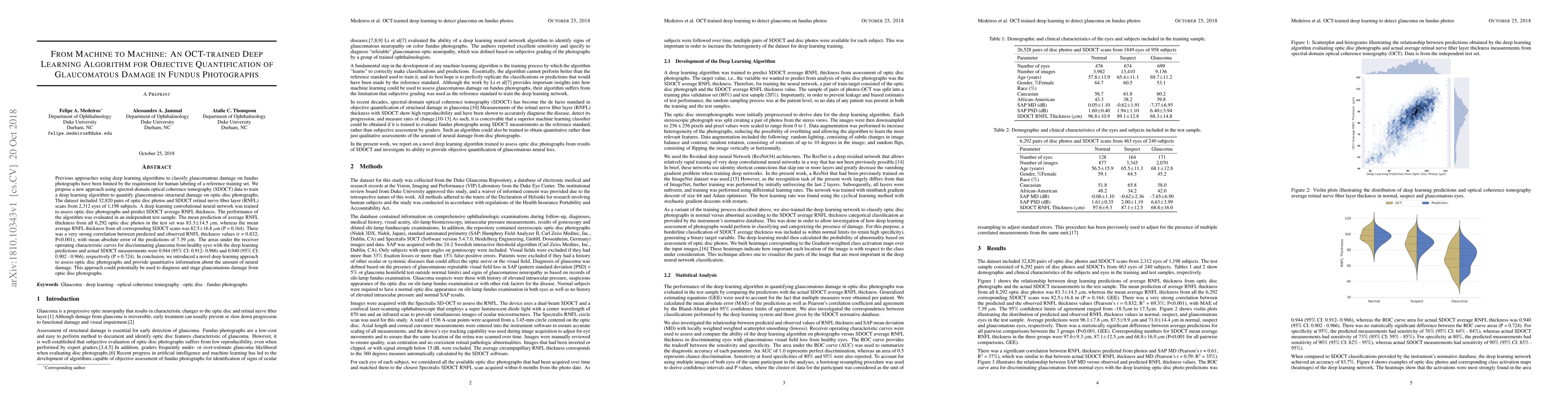
Previous approaches using deep learning algorithms to classify glaucomatous damage on fundus photographs have been limited by the requirement for human labeling of a reference training set. We propose a new approach using spectral-domain optical coherence tomography (SDOCT) data to train a deep learning algorithm to quantify glaucomatous structural damage on optic disc photographs. The dataset included 32,820 pairs of optic disc photos and SDOCT retinal nerve fiber layer (RNFL) scans from 2,312 eyes of 1,198 subjects. A deep learning convolutional neural network was trained to assess optic disc photographs and predict SDOCT average RNFL thickness. The performance of the algorithm was evaluated in an independent test sample. The mean prediction of average RNFL thickness from all 6,292 optic disc photos in the test set was 83.3$\pm$14.5 $\mu$m, whereas the mean average RNFL thickness from all corresponding SDOCT scans was 82.5$\pm$16.8 $\mu$m (P = 0.164). There was a very strong correlation between predicted and observed RNFL thickness values (r = 0.832; P<0.001), with mean absolute error of the predictions of 7.39 $\mu$m. The areas under the receiver operating characteristic curves for discriminating glaucoma from healthy eyes with the deep learning predictions and actual SDOCT measurements were 0.944 (95$\%$ CI: 0.912- 0.966) and 0.940 (95$\%$ CI: 0.902 - 0.966), respectively (P = 0.724). In conclusion, we introduced a novel deep learning approach to assess optic disc photographs and provide quantitative information about the amount of neural damage. This approach could potentially be used to diagnose and stage glaucomatous damage from optic disc photographs.
AI Key Findings
Get AI-generated insights about this paper's methodology, results, and significance.
Paper Details
PDF Preview
Key Terms
Citation Network
Current paper (gray), citations (green), references (blue)
Display is limited for performance on very large graphs.
Similar Papers
Found 4 papersA deep learning approach for automated detection of geographic atrophy from color fundus photographs
APTOS-2024 challenge report: Generation of synthetic 3D OCT images from fundus photographs
Kun Huang, Qiang Chen, Bowen Liu et al.
| Title | Authors | Year | Actions |
|---|
Comments (0)