Full-Field Nanoscale X-ray Diffraction-Contrast Imaging using Direct Detection
Publication
Metrics
AI Quick Summary
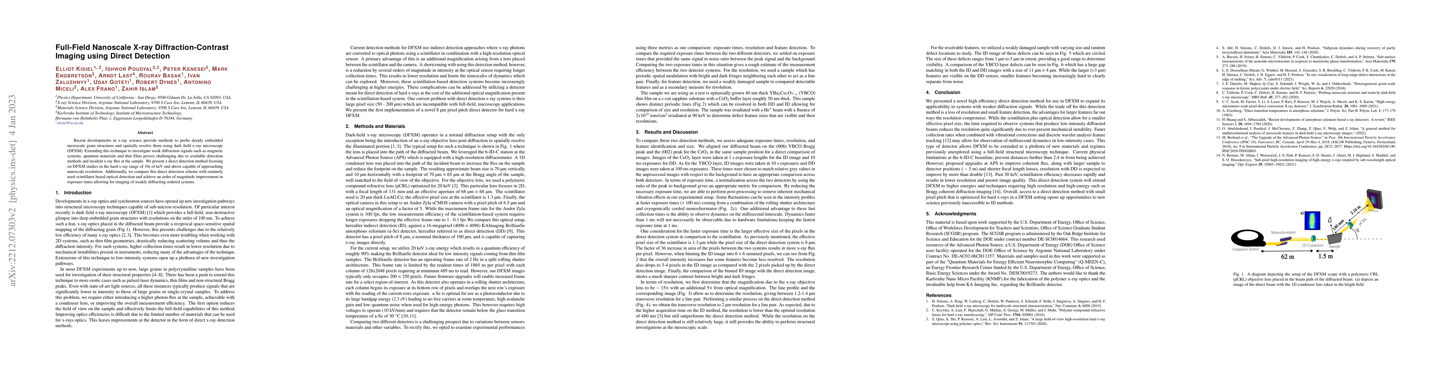
This paper introduces a direct detection method for full-field nanoscale X-ray diffraction-contrast imaging using hard X-rays, significantly improving detection efficiency and reducing exposure times compared to traditional scintillator-based methods, enabling studies of weakly diffracting systems such as magnetic materials and thin films.
Paper Preview
Abstract
Recent developments in x-ray science provide methods to probe deeply embedded mesoscale grain structures and spatially resolve them using dark field x-ray microscopy (DFXM). Extending this technique to investigate weak diffraction signals such as magnetic systems, quantum materials and thin films proves challenging due to available detection methods and incident x-ray flux at the sample. We present a direct detection method focusing on DFXM studies in the hard x-ray range of 10s of keV and above capable of approaching nanoscale resolution. Additionally, we compare this direct detection scheme with routinely used scintillator based optical detection and achieve an order of magnitude improvement in exposure times allowing for imaging of weakly diffracting ordered systems.
AI Key Findings
Get AI-generated insights about this paper's methodology, results, significance, and more — seven facets brought into focus.
Impact
Paper Details
Authors
PDF Preview
Key Terms
Citation Network
Current paper (gray), citations (green), references (blue)
Display is limited for performance on very large graphs.
Discussion 0