Publication
Metrics
AI Quick Summary
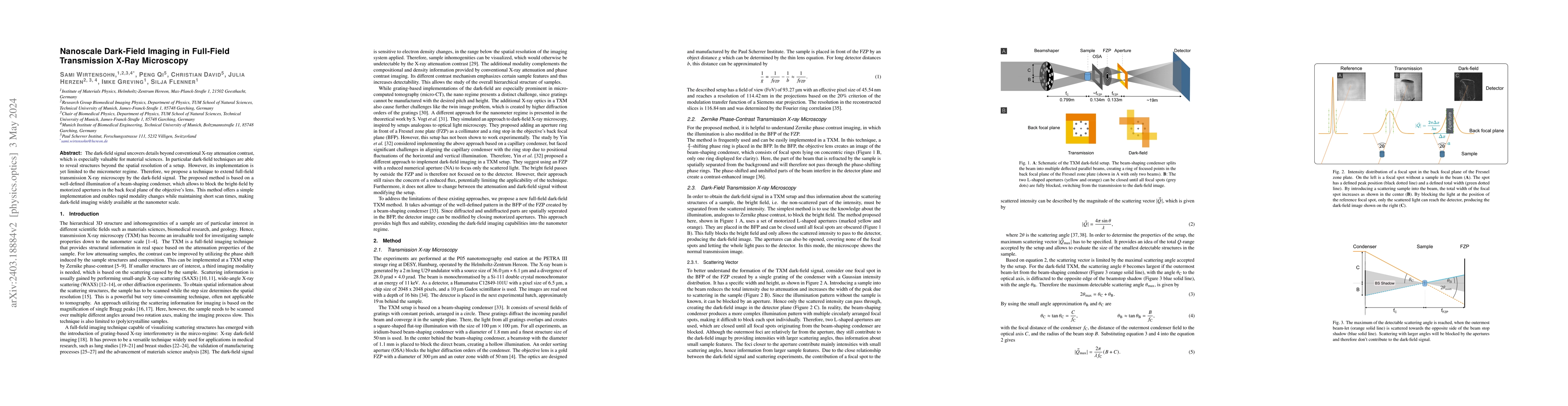
This paper proposes a method to extend full-field transmission X-ray microscopy by incorporating nanoscale dark-field imaging, utilizing a beam-shaping condenser and motorized apertures to block bright-field and reveal finer structural details beyond conventional resolution limits, facilitating rapid modality changes and short scan times.
Paper Preview
Abstract
The dark-field signal uncovers details beyond conventional X-ray attenuation contrast, which is especially valuable for material sciences. In particular, dark-field techniques are able to reveal structures beyond the spatial resolution of a setup. However, its implementation is yet limited to the micrometer regime. Therefore, we propose a technique to extend full-field transmission X-ray microscopy by the dark-field signal. The proposed method is based on a well-defined illumination of a beam-shaping condenser, which allows to block the bright-field by motorized apertures in the back focal plane of the objective's lens. This method offers a simple implementation and enables rapid modality changes while maintaining short scan times, making dark-field imaging widely available at the nanometer scale.
AI Key Findings
Get AI-generated insights about this paper's methodology, results, significance, and more — seven facets brought into focus.
Impact
Paper Details
Authors
PDF Preview
Key Terms
Citation Network
Current paper (gray), citations (green), references (blue)
Display is limited for performance on very large graphs.
Discussion 0