Fully Automated Left Atrium Cavity Segmentation from 3D GE-MRI by Multi-Atlas Selection and Registration
Publication
Metrics
Paper Preview
Abstract
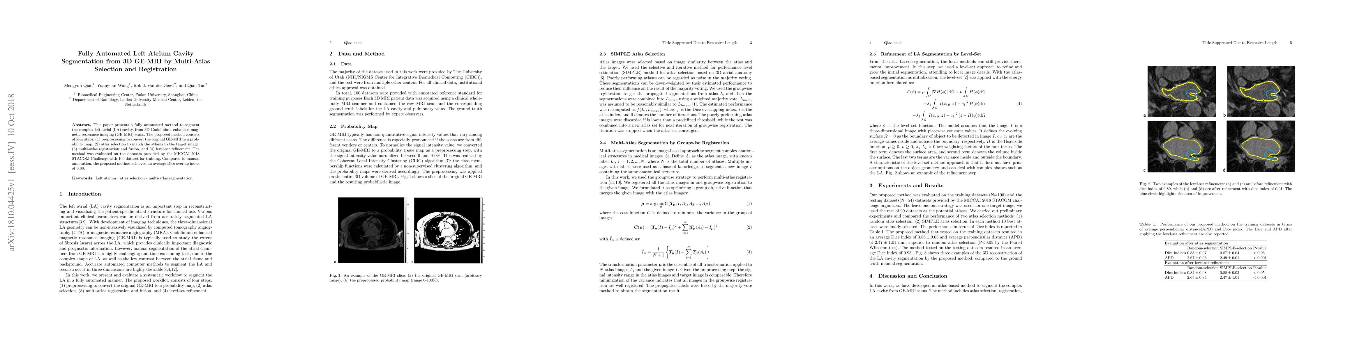
This paper presents a fully automated method to segment the complex left atrial (LA) cavity, from 3D Gadolinium-enhanced magnetic resonance imaging (GE-MRI) scans. The proposed method consists of four steps: (1) preprocessing to convert the original GE-MRI to a probability map, (2) atlas selection to match the atlases to the target image, (3) multi-atlas registration and fusion, and (4) level-set refinement. The method was evaluated on the datasets provided by the MICCAI 2018 STACOM Challenge with 100 dataset for training. Compared to manual annotation, the proposed method achieved an average Dice overlap index of 0.88.
AI Key Findings
Get AI-generated insights about this paper's methodology, results, significance, and more — seven facets brought into focus.
Impact
Paper Details
PDF Preview
Key Terms
Citation Network
Current paper (gray), citations (green), references (blue)
Display is limited for performance on very large graphs.
Discussion 0