Fully Automated Pancreas Segmentation with Two-stage 3D Convolutional Neural Networks
Publication
Metrics
AI Quick Summary
This paper proposes a two-stage 3D convolutional neural network framework for fully automated pancreas segmentation, achieving a mean Dice-Sorensen coefficient of 85.99% on the NIH CT dataset, outperforming other state-of-the-art methods. The first stage uses a U-Net for initial segmentation, followed by a refined segmentation in the second stage on a candidate region.
Paper Preview
Abstract
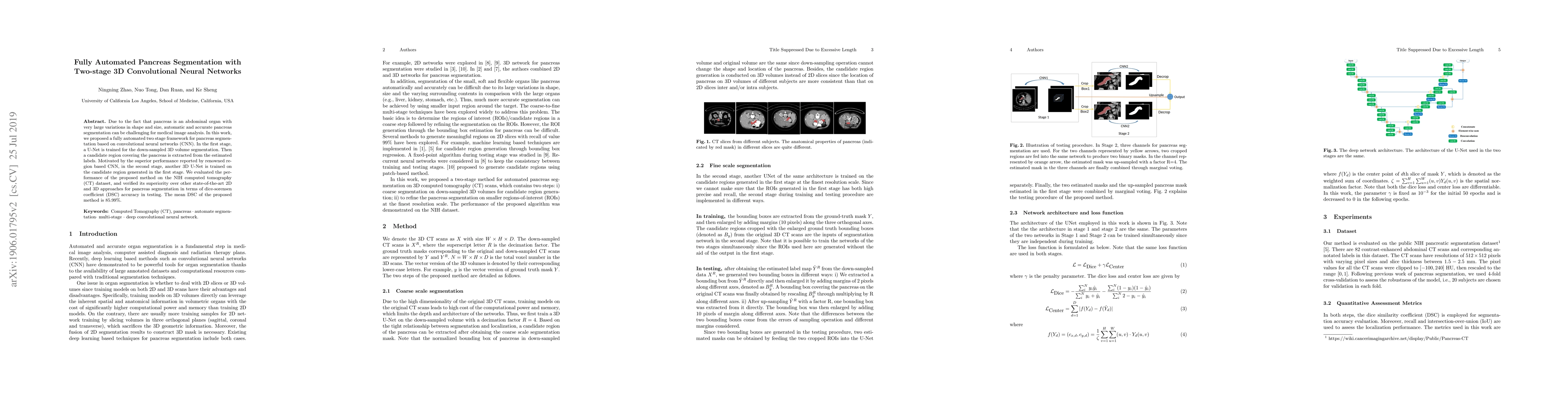
Due to the fact that pancreas is an abdominal organ with very large variations in shape and size, automatic and accurate pancreas segmentation can be challenging for medical image analysis. In this work, we proposed a fully automated two stage framework for pancreas segmentation based on convolutional neural networks (CNN). In the first stage, a U-Net is trained for the down-sampled 3D volume segmentation. Then a candidate region covering the pancreas is extracted from the estimated labels. Motivated by the superior performance reported by renowned region based CNN, in the second stage, another 3D U-Net is trained on the candidate region generated in the first stage. We evaluated the performance of the proposed method on the NIH computed tomography (CT) dataset, and verified its superiority over other state-of-the-art 2D and 3D approaches for pancreas segmentation in terms of dice-sorensen coefficient (DSC) accuracy in testing. The mean DSC of the proposed method is 85.99%.
AI Key Findings
Get AI-generated insights about this paper's methodology, results, significance, and more — seven facets brought into focus.
Impact
Paper Details
PDF Preview
Key Terms
Citation Network
Current paper (gray), citations (green), references (blue)
Display is limited for performance on very large graphs.
Discussion 0