01
MethodologyHow they did it
This study employed 3D fully convolutional neural networks (FCNs) for volumetric segmentation of the pancreas in CT scans.
This paper presents a 3D fully convolutional network for dense volumetric pancreas segmentation in CT scans, achieving state-of-the-art results with an average Dice score of 89.7 ± 3.8% using a summation skip connection architecture. The method is evaluated on a clinical trial dataset of 147 gastric cancer patients.
This study employed 3D fully convolutional neural networks (FCNs) for volumetric segmentation of the pancreas in CT scans. More in Methodology →
Achieved a new state-of-the-art performance with an average Dice score of over 90%. — Demonstrated the effectiveness of multi-GPU training for dense volumetric segmentation tasks. More in Key Results →
This research contributes to the development of automated organ segmentation in medical imaging, with potential applications in cancer diagnosis and treatment. More in Significance →
The dataset used was relatively small compared to other studies. — The proposed method may not generalize well to other organs or imaging modalities. More in Limitations →
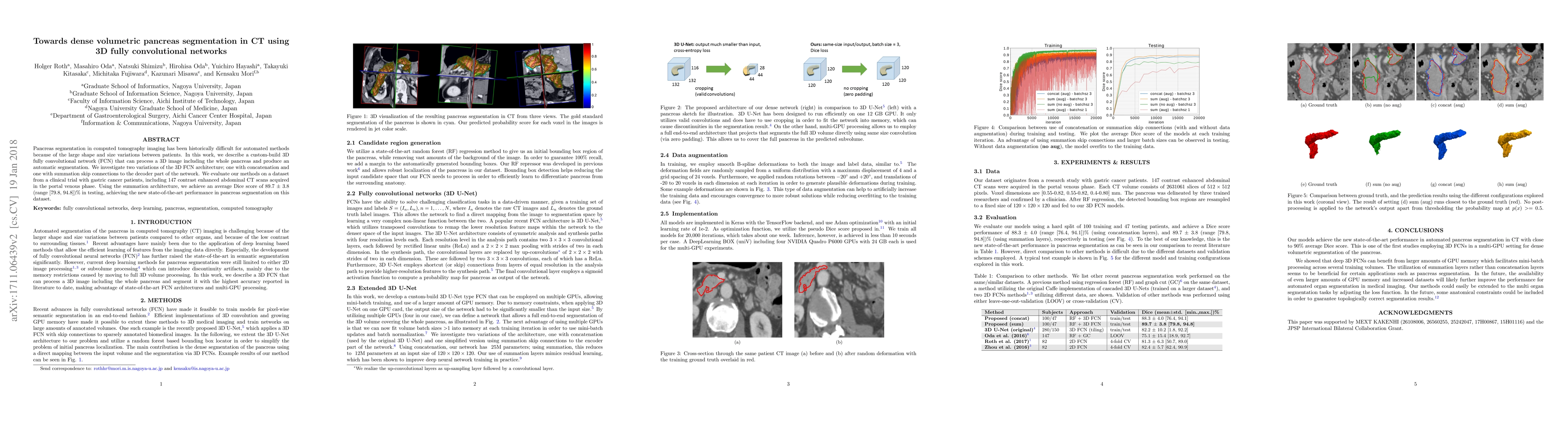
Pancreas segmentation in computed tomography imaging has been historically difficult for automated methods because of the large shape and size variations between patients. In this work, we describe a custom-build 3D fully convolutional network (FCN) that can process a 3D image including the whole pancreas and produce an automatic segmentation. We investigate two variations of the 3D FCN architecture; one with concatenation and one with summation skip connections to the decoder part of the network. We evaluate our methods on a dataset from a clinical trial with gastric cancer patients, including 147 contrast enhanced abdominal CT scans acquired in the portal venous phase. Using the summation architecture, we achieve an average Dice score of 89.7 $\pm$ 3.8 (range [79.8, 94.8]) % in testing, achieving the new state-of-the-art performance in pancreas segmentation on this dataset.
Seven facets of this paper, analysed and brought into focus by AI.
This research contributes to the development of automated organ segmentation in medical imaging, with potential applications in cancer diagnosis and treatment.
This study employed 3D fully convolutional neural networks (FCNs) for volumetric segmentation of the pancreas in CT scans.
This research contributes to the development of automated organ segmentation in medical imaging, with potential applications in cancer diagnosis and treatment.
The development of a novel 3D FCN architecture optimized for dense volumetric segmentation tasks.
This study presents a new approach to automated pancreas segmentation in CT scans, leveraging multi-GPU training and summation layers.
Current paper (gray), citations (green), references (blue)
Display is limited for performance on very large graphs.
Discussion 0