Summary
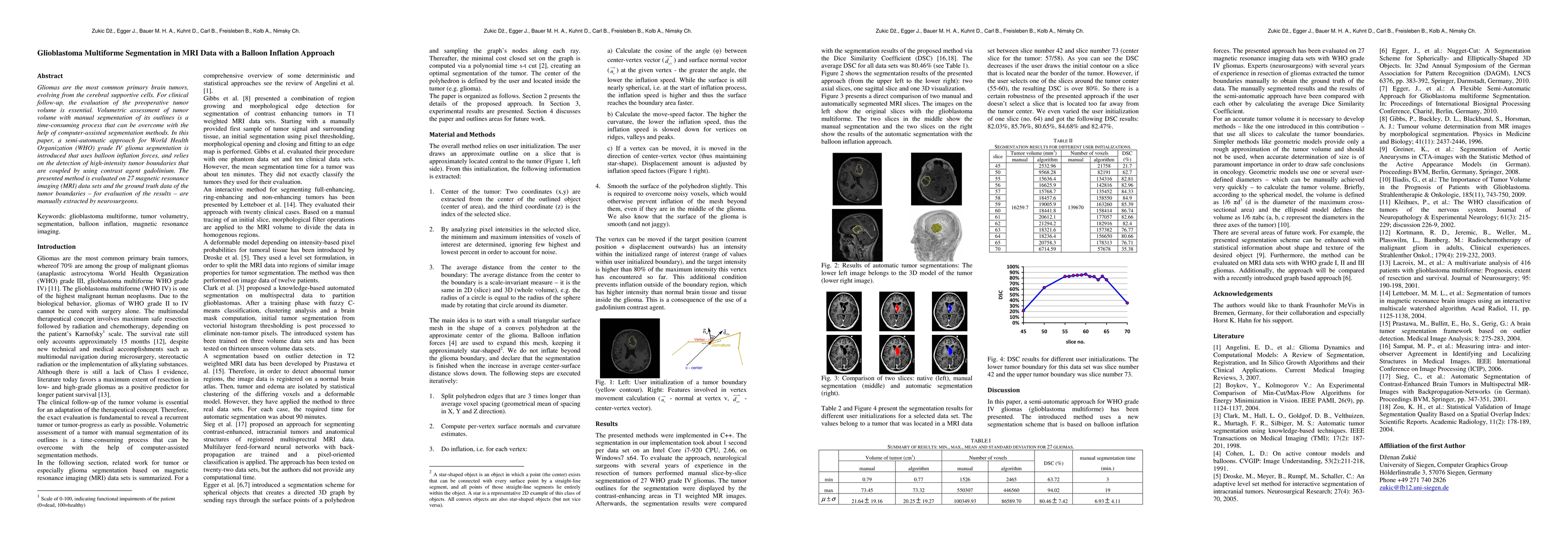
Gliomas are the most common primary brain tumors, evolving from the cerebral supportive cells. For clinical follow-up, the evaluation of the preoperative tumor volume is essential. Volumetric assessment of tumor volume with manual segmentation of its outlines is a time-consuming process that can be overcome with the help of computer-assisted segmentation methods. In this paper, a semi-automatic approach for World Health Organization (WHO) grade IV glioma segmentation is introduced that uses balloon inflation forces, and relies on the detection of high-intensity tumor boundaries that are coupled by using contrast agent gadolinium. The presented method is evaluated on 27 magnetic resonance imaging (MRI) data sets and the ground truth data of the tumor boundaries - for evaluation of the results - are manually extracted by neurosurgeons.
AI Key Findings
Get AI-generated insights about this paper's methodology, results, and significance.
Paper Details
PDF Preview
Key Terms
Citation Network
Current paper (gray), citations (green), references (blue)
Display is limited for performance on very large graphs.
Similar Papers
Found 4 papersRegression and machine learning approaches identify potential risk factors for glioblastoma multiforme.
Felici, Alessio, Peduzzi, Giulia, Pellungrini, Roberto et al.
| Title | Authors | Year | Actions |
|---|

Comments (0)