Publication
Metrics
AI Quick Summary
Gradient optical diffraction tomography (GODT) enables 3D refractive index gradient reconstruction of thick samples, surpassing limitations of traditional ODT. This method uses a coherence-tailored illumination-scanning to achieve high-sensitivity 3D RI gradient imaging, validated both theoretically and experimentally.
Paper Preview
Abstract
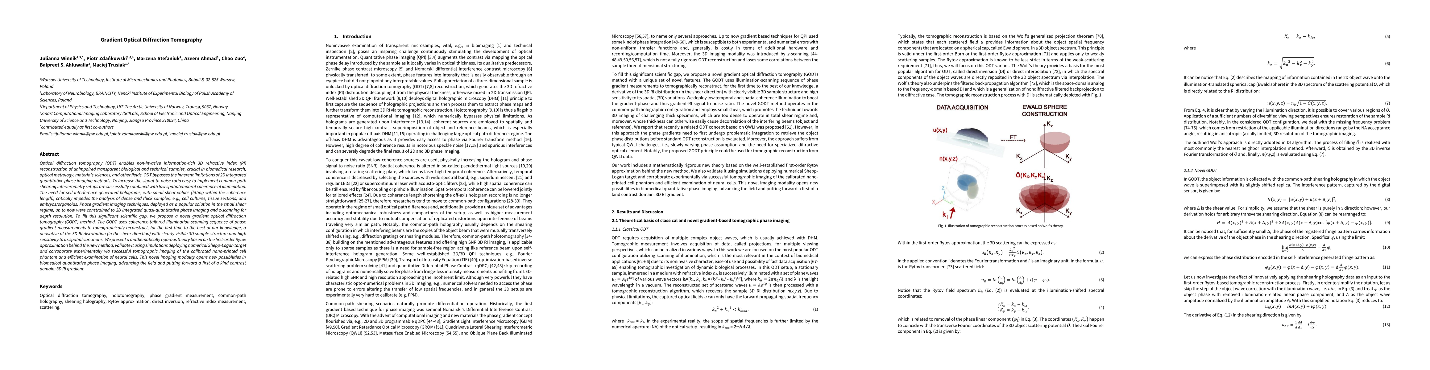
Optical diffraction tomography (ODT) enables non-invasive information-rich 3D refractive index (RI) reconstruction of unimpaired transparent biological and technical samples, crucial in biomedical research, optical metrology, materials sciences, and other fields. ODT bypasses the inherent limitations of 2D integrated quantitative phase imaging methods. To increase the signal-to-noise ratio easy-to-implement common-path shearing interferometry setups are successfully combined with low spatiotemporal coherence of illumination. The need for self-interference generated holograms, with small shear values, critically impedes the analysis of dense and thick samples, e.g., cell cultures, tissue sections, and embryos/organoids. Phase gradient imaging techniques, deployed as a popular solution in the small shear regime, up to now were constrained to 2D integrated quasi-quantitative phase imaging and z-scanning for depth resolution. To fill this significant scientific gap, we propose a novel gradient optical diffraction tomography (GODT) method. The GODT uses coherence-tailored illumination-scanning sequence of phase gradient measurements to tomographically reconstruct, for the first time to the best of our knowledge, a derivative of the 3D RI distribution (in the shear direction) with clearly visible 3D sample structure and high sensitivity to its spatial variations. We present a mathematically rigorous theory based on the first-order Rytov approximation behind the new method, validate it using simulations deploying numerical Shepp-Logan target and corroborate experimentally via successful tomographic imaging of the calibrated nano-printed cell phantom and efficient examination of neural cells. This novel imaging modality opens new possibilities in biomedical quantitative phase imaging, advancing the field and putting forward a first of a kind contrast domain: 3D RI gradient.
AI Key Findings
Get AI-generated insights about this paper's methodology, results, significance, and more — seven facets brought into focus.
Paper Details
Authors
PDF Preview
Related Papers
No references found for this paper.
Discussion 0