01
MethodologyHow they did it
Brief description of the research methodology used
This paper introduces ultra-broadband optical diffraction tomography (ODT), enabling 3D imaging over 150 nm spectral bandwidth with 150 nm resolution. It facilitates precise identification of nanoparticles in cellular structures via spectrally resolved imaging of HeLa cells, offering significant advancements for life and materials science.
This paper introduces ultra-broadband optical diffraction tomography (ODT), enabling 3D imaging over 150 nm spectral bandwidth with 150 nm resolution. It facilitates precise identification of nanoparticles in cellular structures via spectrally resolved imaging of HeLa cells, offering significant advancements for life and materials science.
Brief description of the research methodology used More in Methodology →
Main finding 1 — Main finding 2 More in Key Results →
Why this research is important and its potential impact More in Significance →
Limitation 1 — Limitation 2 More in Limitations →
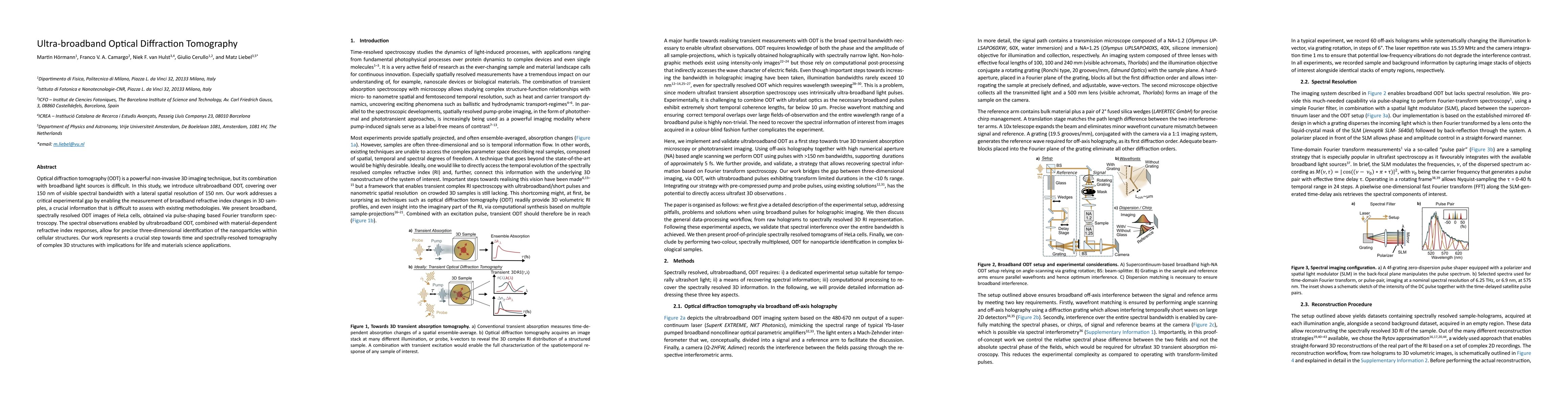
Optical diffraction tomography (ODT) is a powerful non-invasive 3D imaging technique, but its combination with broadband light sources is difficult. In this study, we introduce ultrabroadband ODT, covering over 150 nm of visible spectral bandwidth with a lateral spatial resolution of 150 nm. Our work addresses a critical experimental gap by enabling the measurement of broadband refractive index changes in 3D samples, a crucial information that is difficult to assess with existing methodologies. We present broadband, spectrally resolved ODT images of HeLa cells, obtained via pulse-shaping based Fourier transform spectroscopy. The spectral observations enabled by ultrabroadband ODT, combined with material-dependent refractive index responses, allow for precise three-dimensional identification of the nanoparticles within cellular structures. Our work represents a crucial step towards time and spectrally-resolved tomography of complex 3D structures with implications for life and materials science applications.
Seven facets of this paper, analysed and brought into focus by AI.
Why this research is important and its potential impact
Brief description of the research methodology used
Why this research is important and its potential impact
Main technical or theoretical contribution
What makes this work novel or different from existing research
Current paper (gray), citations (green), references (blue)
Display is limited for performance on very large graphs.
Discussion 0