Publication
Metrics
AI Quick Summary
This paper demonstrates imaging of a 210 nm graphene sheet with 2 Å resolution using coherent diffraction and holography with low-energy electrons, reconstructing the entire sheet from a single diffraction pattern. The method's potential for non-damaging imaging of biological molecules opens avenues for protein deposition on graphene sheets.
Paper Preview
Abstract
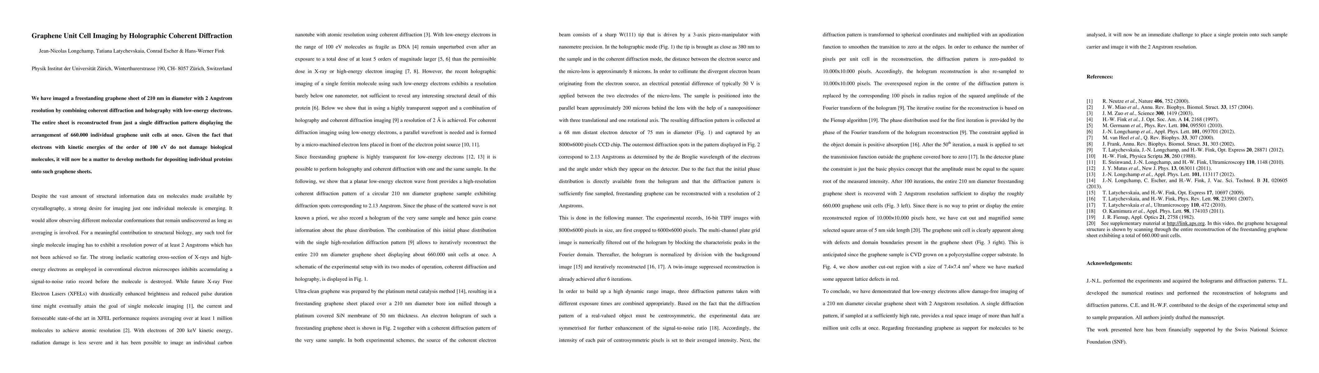
We have imaged a freestanding graphene sheet of 210 nm in diameter with 2 Angstrom resolution by combining coherent diffraction and holography with low-energy electrons. The entire sheet is reconstructed from just a single diffraction pattern displaying the arrangement of 660.000 individual graphene unit cells at once. Given the fact that electrons with kinetic energies of the order of 100 eV do not damage biological molecules, it will now be a matter to develop methods for depositing individual proteins onto such graphene sheets.
AI Key Findings
Get AI-generated insights about this paper's methodology, results, significance, and more — seven facets brought into focus.
Impact
Paper Details
PDF Preview
Key Terms
Citation Network
Current paper (gray), citations (green), references (blue)
Display is limited for performance on very large graphs.
Discussion 0