Publication
Metrics
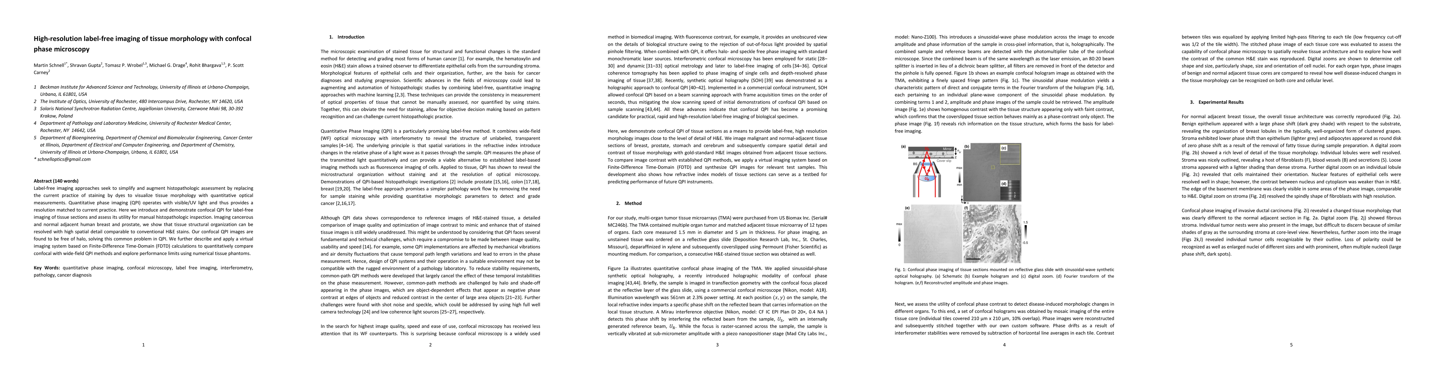
AI Quick Summary
This paper introduces confocal quantitative phase imaging (QPI) for label-free tissue morphology visualization, demonstrating high-resolution imaging of human breast and prostate tissue comparable to traditional H&E stains. The method resolves structural organization without the halo effect common in QPI, and a virtual imaging system based on FDTD calculations compares its performance with wide-field QPI.
Paper Preview
Abstract
Label-free imaging approaches seek to simplify and augment histopathologic assessment by replacing the current practice of staining by dyes to visualize tissue morphology with quantitative optical measurements. Quantitative phase imaging (QPI) operates with visible/UV light and thus provides a resolution matched to current practice. Here we introduce and demonstrate confocal QPI for label-free imaging of tissue sections and assess its utility for manual histopathologic inspection. Imaging cancerous and normal adjacent human breast and prostate, we show that tissue structural organization can be resolved with high spatial detail comparable to conventional H&E stains. Our confocal QPI images are found to be free of halo, solving this common problem in QPI. We further describe and apply a virtual imaging system based on Finite-Difference Time-Domain (FDTD) calculations to quantitatively compare confocal with wide-field QPI methods and explore performance limits using numerical tissue phantoms.
AI Key Findings
Get AI-generated insights about this paper's methodology, results, significance, and more — seven facets brought into focus.
Impact
Paper Details
Authors
PDF Preview
Key Terms
Citation Network
Current paper (gray), citations (green), references (blue)
Display is limited for performance on very large graphs.
Discussion 0