This paper presents a holographic intravital microscopy technique for quantitative, label-free imaging of circulating blood cells in live mice, providing 2-D and 3-D morphological and biochemical information, including cell shape, haemoglobin concentration, and contents. The method is applied to study blood flow dynamics in healthy and sepsis-model mice.
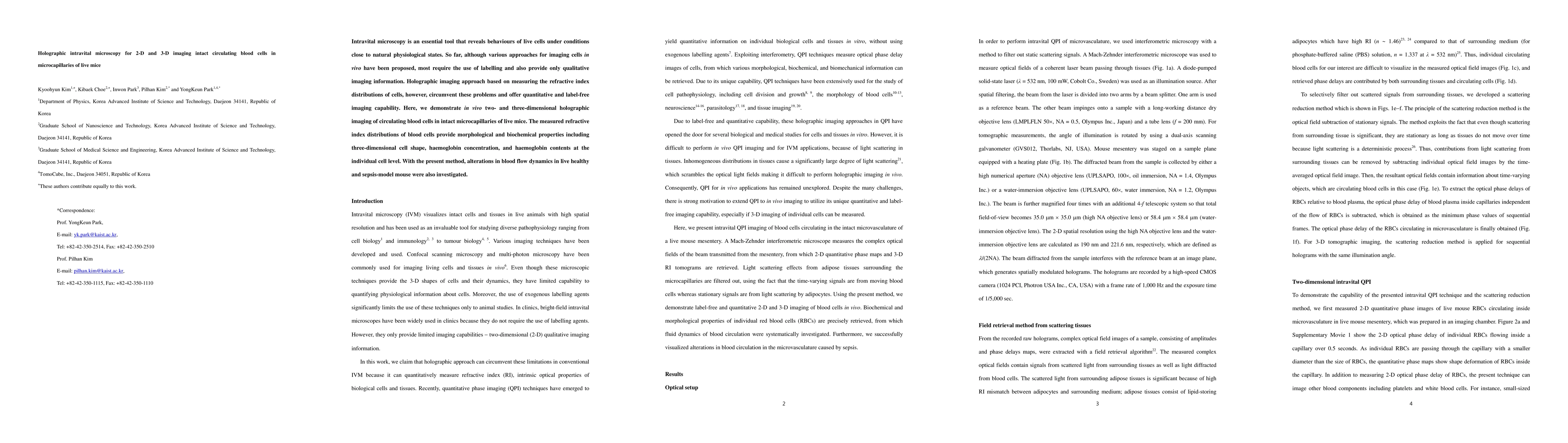
Intravital microscopy is an essential tool that reveals behaviours of live cells under conditions close to natural physiological states. So far, although various approaches for imaging cells in vivo have been proposed, most require the use of labelling and also provide only qualitative imaging information. Holographic imaging approach based on measuring the refractive index distributions of cells, however, circumvent these problems and offer quantitative and label-free imaging capability. Here, we demonstrate in vivo two- and three-dimensional holographic imaging of circulating blood cells in intact microcapillaries of live mice. The measured refractive index distributions of blood cells provide morphological and biochemical properties including three-dimensional cell shape, haemoglobin concentration, and haemoglobin contents at the individual cell level. With the present method, alterations in blood flow dynamics in live healthy and sepsis-model mouse were also investigated.
This graph shows how this paper is connected to other research. The current paper is shown in dark gray, papers that cite it are in green, and papers it references are in blue. You can:
• Hover over any node to see the paper title
• Click a node to view that paper's details
• Drag nodes to rearrange the graph
• Use the zoom controls (bottom right) to zoom in/out
Title | Authors | Year | Actions |
---|---|---|---|
Novel imaging and biophysical approaches to study tissue hydraulics in mammalian folliculogenesis
|
Jake Turley, Kim Whye Leong, C. J. Chan
|
2024
|
View Paper |
3D Bioprinting Cell Detection Using Autofocused Compressive Holography
|
Zhou Ge, Jingyuan Zhu, Aoxiang Jin, Yuanyuan Liu, Wenjing Zhou, Yingjie Yu
|
2024
|
View Paper |
Changes to the shape, orientation and packing of red cells as a function of retinal capillary size
|
P. Bedggood, Yifu Ding, A. Metha
|
2023
|
View Paper |
Analysis of structural effects of sickle cell disease on brain vasculature of mice using three-dimensional quantitative phase imaging
|
Caroline Filan, Hannah Song, Manu O Platt, Francisco E. Robles
|
2023
|
View Paper |
Optical diffraction tomography and image reconstruction to measure host cell alterations caused by divergent Plasmodium species.
|
J. J. Ong, Jeonghun Oh, Xiang Yong Ang, Renugah Naidu, T. Chu, Jae Hyoung Im, Umar Manzoor, Tuyet Kha Nguyen, Seok-Won Na, E. Han, Christeen Davis, Won Sun Park, W. Chun, Hojong Jun, Se Jin Lee, Sunghun Na, J. Chan, YongKeun Park, B. Russell, Rajesh Chandramohanadas, Jin-Hee Han
|
2022
|
View Paper |
Quantitative Phase Imaging: Recent Advances and Expanding Potential in Biomedicine.
|
Thang L Nguyen, Soorya Pradeep, Robert Judson-Torres, J. Reed, M. Teitell, T. Zangle
|
2022
|
View Paper |
Intravital Laser-scanning Two-photon and Confocal Microscopy for Biomedical Research
|
Jieun Moon, P. Kim
|
2021
|
View Paper |
Quantitative blood flow estimation in vivo by optical speckle image velocimetry
|
Muhammad Mohsin Qureshi, Yan Liu, K. D. Mac, Minsung Kim, Abdul Mohaimen Safi, Euiheon Chung
|
2021
|
View Paper |
Measurement of Cell Volume Using In-Line Digital Holography.
|
Hangjian Ling, K. Sridhar, Sumanth Gollapudi, Jyoti Kumar, R. Ohgami
|
2020
|
View Paper |
Deep learning-based digital in-line holographic microscopy for high resolution with extended field of view
|
Hyeokjun Byeon, Taesik Go, S. Lee
|
2019
|
View Paper |
Cycle-consistent deep learning approach to coherent noise reduction in optical diffraction tomography.
|
Gunho Choi, DongHun Ryu, YoungJu Jo, Youngseo Kim, W. Park, Hyun-Seok Min, Yongkeun Park
|
2018
|
View Paper |
Mechanical Mapping of Spinal Cord Growth and Repair in Living Zebrafish Larvae by Brillouin Imaging
|
Raimund Schlüßler, Stephanie Möllmert, Shada Abuhattum, G. Cojoc, Paul Müller, Kyoohyun Kim, Conrad Möckel, C. Zimmermann, J. Czarske, J. Guck
|
2018
|
View Paper |
Endoscopic diffraction phase microscopy.
|
Chenfei Hu, Shuaishuai Zhu, Liang Gao, G. Popescu
|
2018
|
View Paper |
Digital stereo-holographic microscopy for studying three-dimensional particle dynamics
|
Hyeokjun Byeon, Taesik Go, S. Lee
|
2018
|
View Paper |
Intravital imaging of a pulmonary endothelial surface layer in a murine sepsis model.
|
I. Park, K. Choe, H. Seo, Y. Hwang, E. Song, Jinhyo Ahn, You Hwan Jo, Pilhan Kim
|
2018
|
View Paper |
Quantitative phase microscopy for evaluation of intestinal inflammation and wound healing utilizing label-free biophysical markers.
|
D. Bettenworth, A. Bokemeyer, C. Poremba, N. Ding, S. Ketelhut, P. Lenz, B. Kemper
|
2018
|
View Paper |
Correlative three-dimensional fluorescence and refractive index tomography: bridging the gap between molecular specificity and quantitative bioimaging.
|
Kyoohyun Kim, W. Park, Sangchan Na, Sangbum Kim, Taehong Kim, Won Do Heo, Yongkeun Park
|
2017
|
View Paper |
Numerical-experimental observation of shape bistability of red blood cells flowing in a microchannel.
|
A. Guckenberger, Alexander Kihm, Thomas John, C. Wagner, S. Gekle
|
2017
|
View Paper |
Digital Holography, a metrological tool for quantitative analysis: Trends and future applications
|
M. Paturzo, V. Pagliarulo, V. Bianco, P. Memmolo, L. Miccio, F. Merola, P. Ferraro
|
2017
|
View Paper |
Measurements of three-dimensional refractive index tomography and membrane deformability of live erythrocytes from Pelophylax nigromaculatus
|
Geon Kim, Moosung Lee, SeongYeon Youn, EuiTae Lee, Daeheon Kwon, Jonghun Shin, Sangyun Lee, Youn Sil Lee, Yongkeun Park
|
2017
|
View Paper |
High-speed video capillaroscopy method for imaging and evaluation of moving red blood cells
|
I. Gurov, M. Volkov, N. Margaryants, A. Pimenov, A. Potemkin
|
2017
|
View Paper |
Correlative three-dimensional fluorescence and refractive index tomography: bridging the gap between molecular specificity and quantitative bioimaging
|
Kyoohyun Kim, W. Park, Sangchan Na, Sangbum Kim, Taehong Kim, W. Heo, Yongkeun Park
|
2017
|
View Paper |
Mechanical mapping of spinal cord development and repair in living zebrafish larvae using Brillouin microscopy
|
Raimund Schlüβler, Stephanie Möllmert, Shada Abuhattum, Conrad Möckel, C. Zimmermann, J. Czarske, J. Guck
|
2017
|
View Paper |
Identification of non-activated lymphocytes using three-dimensional refractive index tomography and machine learning
|
Jonghee Yoon, YoungJu Jo, Min-hyeok Kim, Kyoohyun Kim, Sangyun Lee, Suk-Jo Kang, Yongkeun Park
|
2017
|
View Paper |
Measurements of morphological and biophysical alterations in individual neuron cells associated with early neurotoxic effects in Parkinson's disease
|
Su-A Yang, Jonghee Yoon, Kyoohyun Kim, Yongkeun Park
|
2017
|
View Paper |
Tomographic phase microscopy: principles and applications in bioimaging [Invited].
|
Di Jin, R. Zhou, Z. Yaqoob, P. So
|
2017
|
View Paper |
Rapid in‐focus corrections on quantitative amplitude and phase imaging using transport of intensity equation method
|
Xin Meng, Xiaolin Tian, Yan Kong, Aihui Sun, Wei Yu, Weiying Qian, Xiaojun Song, Haoyang Cui, Liang Xue, Cheng Liu, Shouyu Wang
|
2017
|
View Paper |
Label-free identification of non-activated lymphocytes using three-dimensional refractive index tomography and machine learning
|
Jonghee Yoon, YoungJu Jo, Min-hyeok Kim, Kyoohyun Kim, Sangyun Lee, Suk-Jo Kang, Yongkeun Park
|
2017
|
View Paper |
Holotomography: refractive index as an intrinsic imaging contrast for 3-D label-free live cell imaging
|
Doyeon Kim, Sangyun Lee, Moosung Lee, Juntaek Oh, Su-A Yang, Yongkeun Park
|
2017
|
View Paper |
Time-multiplexed structured illumination using a DMD for optical diffraction tomography.
|
KyeoReh Lee, Kyoohyun Kim, Geon Kim, Seungwoo Shin, Yongkeun Park
|
2016
|
View Paper |
Measurements of morphological and biochemical alterations in individual neuron cells associated with early neurotoxic effects in Parkinson’s disease
|
Su-A Yang, Jonghee Yoon, Kyoohyun Kim, Yongkeun Park
|
2016
|
View Paper |
Three-dimensional imaging through thick phase-fluctuating medium based on phase-shift digital holography with two adjacent light sources for common-path geometry
|
Shutaro Kodama, M. Ohta, Y. Miyamoto, W. Osten, Mitsuo Takeda, E. Watanabe
|
2021
|
View Paper |
Holographic microscopy for biomedical imaging.
|
D. Donnarumma
|
2018
|
View Paper |
An Introduction to Lensless Digital Holographic Microscopy
|
N. Lindquist
|
2018
|
View Paper |
Correlative three-dimensional fluorescence and refractive index tomography : bridging the gap between molecular specificity and quantitative bioimaging
|
YooHyun, Im, Angchan, ong, Eun, Ark
|
2017
|
View Paper |
Published: 2019-01-16
Published: 2021-06-15
Comments (0)