Publication
Metrics
Paper Preview
Abstract
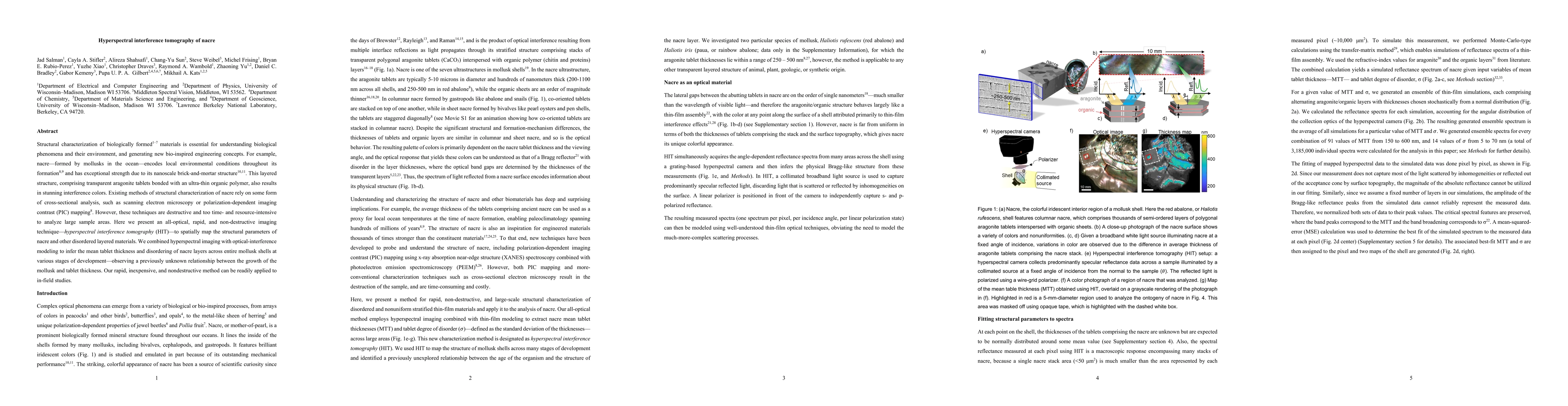
Structural characterization of biologically formed materials is essential for understanding biological phenomena and their environment, and generating new bio-inspired engineering concepts. For example, nacre -- formed by mollusks in the ocean -- encodes local environmental conditions throughout its formation and has exceptional strength due to its nanoscale brick-and-mortar structure. This layered structure, comprising transparent aragonite tablets bonded with an ultra-thin organic polymer, also results in stunning interference colors. Existing methods of structural characterization of nacre rely on some form of cross-sectional analysis, such as scanning electron microscopy or polarization-dependent imaging contrast (PIC) mapping. However, these techniques are destructive and too time- and resource-intensive to analyze large sample areas. Here we present an all-optical, rapid, and non-destructive imaging technique -- hyperspectral interference tomography (HIT) -- to spatially map the structural parameters of nacre and other disordered layered materials. We combined hyperspectral imaging with optical-interference modeling to infer the mean tablet thickness and disordering of nacre layers across entire mollusk shells at various stages of development, observing a previously unknown relationship between the growth of the mollusk and tablet thickness. Our rapid, inexpensive, and nondestructive method can be readily applied to in-field studies.
AI Key Findings
Get AI-generated insights about this paper's methodology, results, significance, and more — seven facets brought into focus.
Impact
Paper Details
Authors
PDF Preview
Key Terms
Citation Network
Current paper (gray), citations (green), references (blue)
Display is limited for performance on very large graphs.
Discussion 0