Publication
Metrics
AI Quick Summary
A new technique using hyperspectral light allows for non-invasive 3D imaging of refractive indices in biological samples at the microscopic level, with applications for precise molecular analysis.
Paper Preview
Abstract
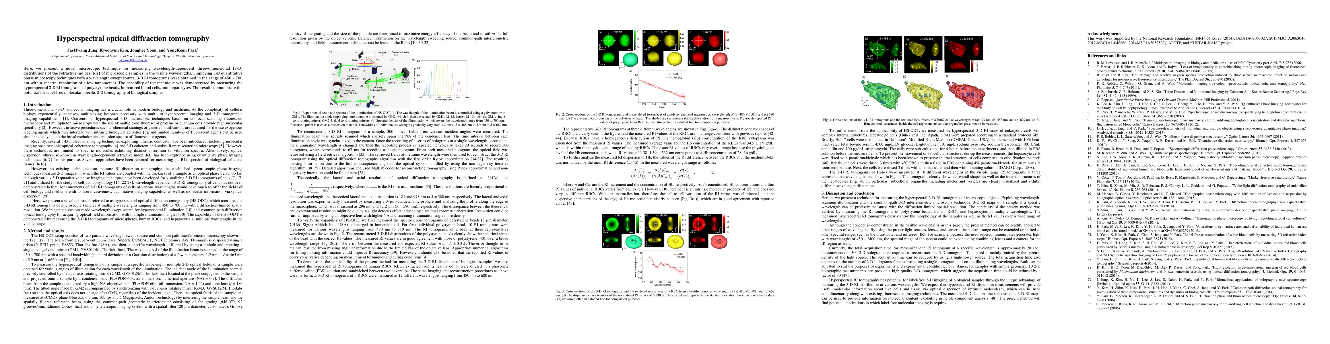
Here, we present a novel microscopic technique for measuring wavelength-dependent three-dimensional (3-D) distributions of the refractive indices (RIs) of microscopic samples in the visible wavelengths. Employing 3-D quantitative phase microscopy techniques with a wavelength-swept source, 3-D RI tomograms were obtained in the range of 450 - 700 nm with a spectral resolution of a few nanometers. The capability of the technique was demonstrated by measuring the hyperspectral 3-D RI tomograms of polystyrene beads, human red blood cells, and hepatocytes. The results demonstrate the potential for label-free molecular specific 3-D tomography of biological samples.
AI Key Findings
Get AI-generated insights about this paper's methodology, results, significance, and more — seven facets brought into focus.
Impact
Paper Details
PDF Preview
Key Terms
Citation Network
Current paper (gray), citations (green), references (blue)
Display is limited for performance on very large graphs.
Discussion 0