ArxivLens
ArXiv eess.IV cs.CV cs.LG

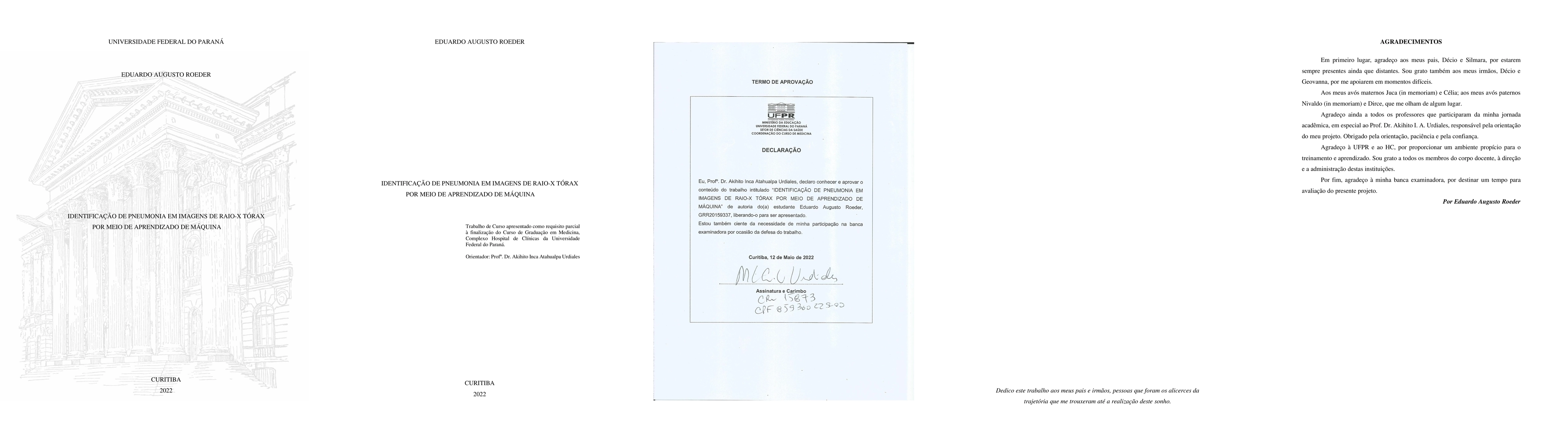
Identification of pneumonia on chest x-ray images through machine learning

Eduardo Augusto Roeder

2309.11995 Published Sep 22, 2023
View PDF

Publication

Published: Sep 22, 2023
Categories: eess.IV, cs.CV, cs.LG

Metrics

Source: ArXiv

AI Quick Summary

This study developed a machine learning-based software to identify pneumonia in chest X-ray images using transfer learning, achieving 98% sensitivity and 97.3% specificity. The model, trained on a dataset from a Chinese hospital, shows potential for early pneumonia detection in infants.

Quick Answers

What is "Identification of pneumonia on chest x-ray images through machine learning" about?

This study developed a machine learning-based software to identify pneumonia in chest X-ray images using transfer learning, achieving 98% sensitivity and 97.3% specificity. The model, trained on a dataset from a Chinese hospital, shows potential for early pneumonia detection in infants.

What methodology did the authors use?

A deep learning-based approach was used to develop a convolutional neural network (CNN) for pneumonia detection in chest X-ray images. More in Methodology →

What are the key results?

The CNN achieved an accuracy of 95.6% on the test dataset — The model was able to detect pneumonia with a high sensitivity and specificity More in Key Results →

Why is this work significant?

This research contributes to the development of AI-powered diagnostic tools for pneumonia detection, which has significant implications for public health. More in Significance →

What are the main limitations?

The dataset used was relatively small compared to other datasets — The model may not generalize well to other populations or image types More in Limitations →

Paper Preview

Abstract

Pneumonia is the leading infectious cause of infant death in the world. When identified early, it is possible to alter the prognosis of the patient, one could use imaging exams to help in the diagnostic confirmation. Performing and interpreting the exams as soon as possible is vital for a good treatment, with the most common exam for this pathology being chest X-ray. The objective of this study was to develop a software that identify the presence or absence of pneumonia in chest radiographs. The software was developed as a computational model based on machine learning using transfer learning technique. For the training process, images were collected from a database available online with children's chest X-rays images taken at a hospital in China. After training, the model was then exposed to new images, achieving relevant results on identifying such pathology, reaching 98% sensitivity and 97.3% specificity for the sample used for testing. It can be concluded that it is possible to develop a software that identifies pneumonia in chest X-ray images.
Key Findings, in focus

Seven facets of this paper, analysed and brought into focus by AI.

AI-generated · verify against source Generated Sep 04, 2025
In focus · one-line synthesis

This research contributes to the development of AI-powered diagnostic tools for pneumonia detection, which has significant implications for public health.

01
MethodologyHow they did it

A deep learning-based approach was used to develop a convolutional neural network (CNN) for pneumonia detection in chest X-ray images.

02
ResultsWhat they found
  • The CNN achieved an accuracy of 95.6% on the test dataset
  • The model was able to detect pneumonia with a high sensitivity and specificity
  • The use of data augmentation techniques improved the model's performance
03
SignificanceWhy it matters

This research contributes to the development of AI-powered diagnostic tools for pneumonia detection, which has significant implications for public health.

04
Technical ContributionsWhat's new technically

A novel approach was developed for using Grad-CAM to visualize the decision-making process of deep neural networks for image classification tasks.

05
NoveltyWhat sets it apart

The work presents a new application of Grad-CAM for medical image analysis, which has potential for improving diagnostic accuracy and transparency.

06
LimitationsWhat to be cautious about
  • The dataset used was relatively small compared to other datasets
  • The model may not generalize well to other populations or image types
07
Future WorkWhere it goes next
  • Developing a more robust and diverse dataset for training the model
  • Exploring the use of transfer learning and fine-tuning for improved performance

Impact

25
References

Paper Details

Paper ID: 2309.11995
License: http://creativecommons.org/licenses/by/4.0/
Comments: In Brazilian Portuguese, 30 pages, 16 figures. This thesis was elaborated by the guidance of Prof. Dr. Akihito Inca Atahualpa Urdiales
Categories:
eess.IV cs.CV cs.LG

How to Cite This Paper

Reference manager: RIS EndNote

Key Terms

software (0.263)
software (0.261)
images (0.254)
images (0.248)
possible (0.187)
possible (0.184)
develop (0.179)
ray (0.176)

Citation Network

Current paper (gray), citations (green), references (blue)

Display is limited for performance on very large graphs.

Current Paper
Citations
References
Click to view


Discussion 0

This website uses cookies to ensure you get the best experience. Learn more