Pneumonia Detection on Chest X-ray using Radiomic Features and Contrastive Learning
Publication
Metrics
AI Quick Summary
This paper proposes a framework combining radiomics features and contrastive learning to detect pneumonia in chest X-rays, achieving superior performance compared to state-of-the-art models and enhancing model interpretability. The method demonstrates significant improvements in F1-score on the RSNA Pneumonia Detection Challenge dataset.
Paper Preview
Abstract
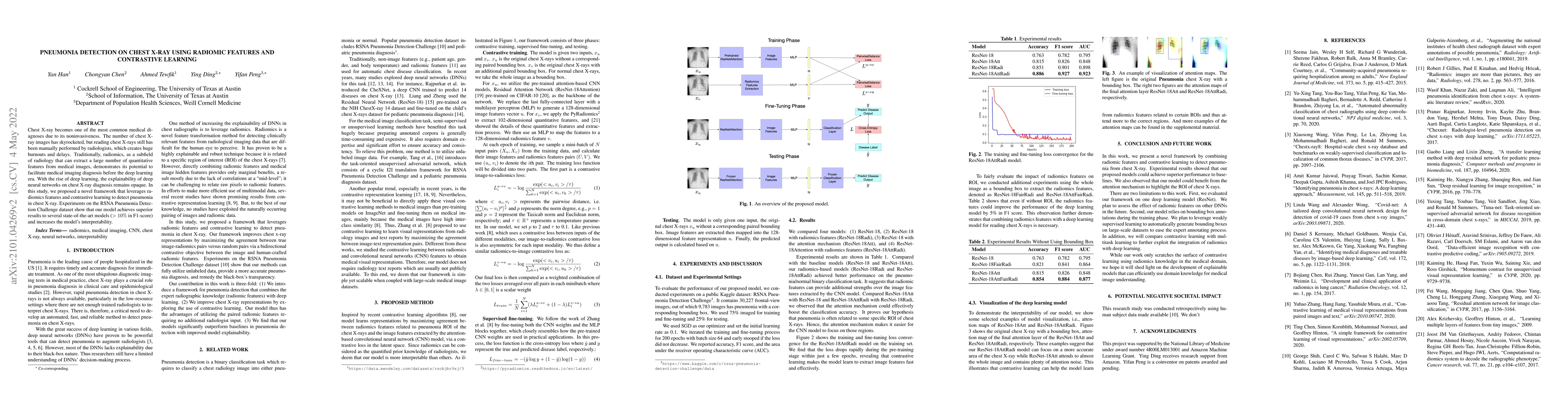
Chest X-ray becomes one of the most common medical diagnoses due to its noninvasiveness. The number of chest X-ray images has skyrocketed, but reading chest X-rays still have been manually performed by radiologists, which creates huge burnouts and delays. Traditionally, radiomics, as a subfield of radiology that can extract a large number of quantitative features from medical images, demonstrates its potential to facilitate medical imaging diagnosis before the deep learning era. With the rise of deep learning, the explainability of deep neural networks on chest X-ray diagnosis remains opaque. In this study, we proposed a novel framework that leverages radiomics features and contrastive learning to detect pneumonia in chest X-ray. Experiments on the RSNA Pneumonia Detection Challenge dataset show that our model achieves superior results to several state-of-the-art models (> 10% in F1-score) and increases the model's interpretability.
AI Key Findings
Get AI-generated insights about this paper's methodology, results, significance, and more — seven facets brought into focus.
Impact
Paper Details
Authors
PDF Preview
Key Terms
Citation Network
Current paper (gray), citations (green), references (blue)
Display is limited for performance on very large graphs.
Discussion 0