Summary
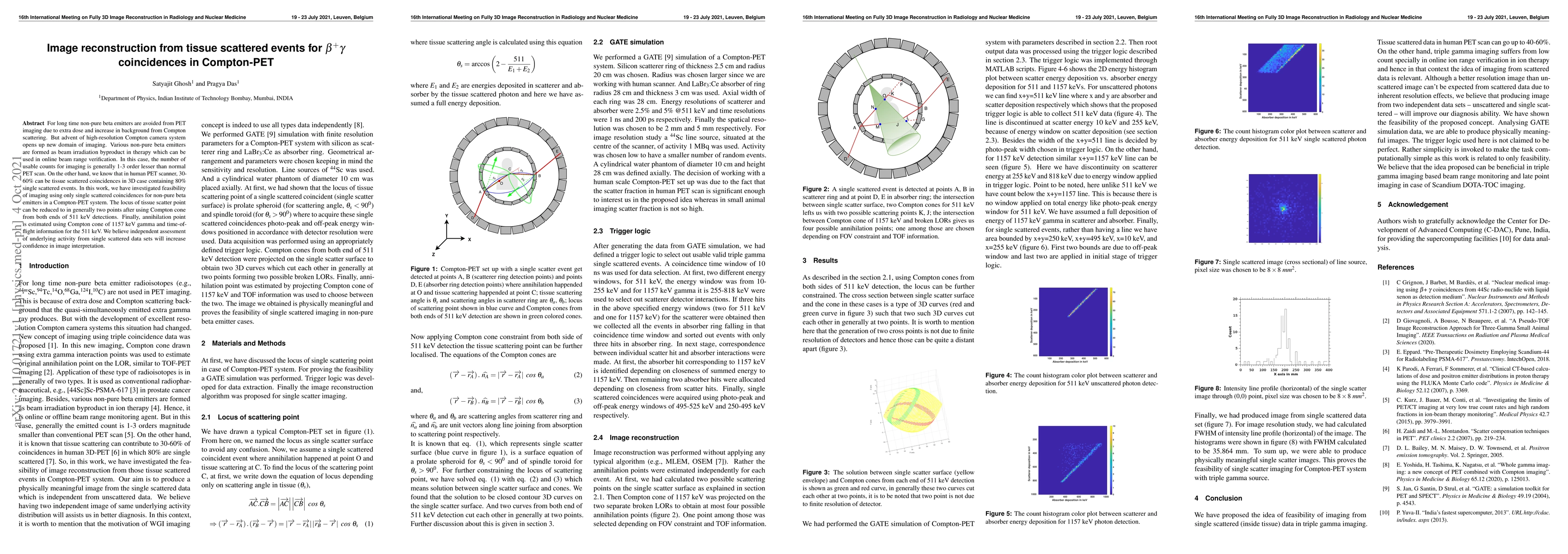
For long time non-pure beta emitters are avoided from PET imaging due to extra dose and increase in background from Compton scattering. But advent of high-resolution Compton camera system opens up new domain of imaging. Various non-pure beta emitters are formed as beam irradiation byproduct in therapy which can be used in online beam range verification. In this case, the number of usable counts for imaging is generally 1-3 order lesser than normal PET scan. On the other hand, we know that in human PET scanner, 30-60\% can be tissue scattered coincidences in 3D case containing 80\% single scattered events. In this work, we have investigated feasibility of imaging using only single scattered coincidences for non-pure beta emitters in a Compton-PET system. The locus of tissue scatter point can be reduced to in generally two points after using Compton cone from both ends of 511 keV detections. Finally, annihilation point is estimated using Compton cone of 1157 keV gamma and time-of-flight information for the 511 keV. We believe independent assessment of underlying activity from single scattered data sets will increase confidence in image interpretation.
AI Key Findings
Get AI-generated insights about this paper's methodology, results, and significance.
Paper Details
PDF Preview
Key Terms
Citation Network
Current paper (gray), citations (green), references (blue)
Display is limited for performance on very large graphs.
Similar Papers
Found 4 papersDirect3γ: A Pipeline for Direct Three-gamma PET Image Reconstruction
Alexandre Bousse, Dimitris Visvikis, Youness Mellak et al.
Reconstruction of multiple Compton scattering events in MeV gamma-ray Compton telescopes towards GRAMS: the physics-based probabilistic model
Masato Kimura, Yoshiyuki Inoue, Hirokazu Odaka et al.
No citations found for this paper.

Comments (0)