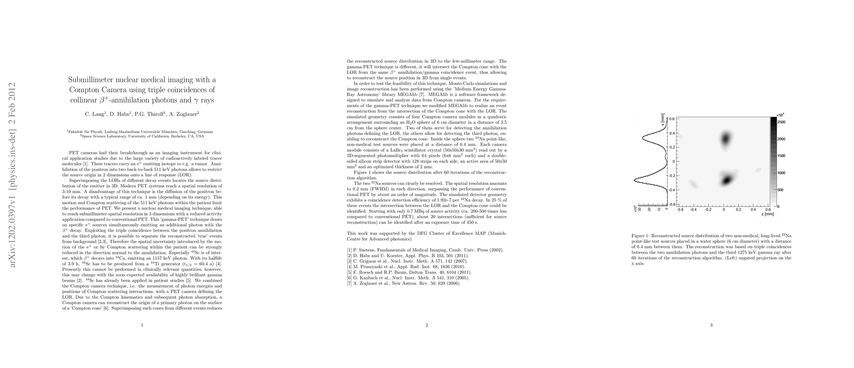
Summary
Modern PET systems reach a spatial resolution of 3-10 mm. A disadvantage of this technique is the diffusion of the positron before its decay with a typical range of ca. 1 mm (depending on its energy). This motion and Compton scattering of the 511 keV photons within the patient limit the performance of PET. We present a nuclear medical imaging technique, able to reach submillimeter spatial resolution in 3 dimensions with a reduced activity application compared to conventional PET. This 'gamma-PET' technique draws on specific positron sources simultaneously emitting an additional photon with the \beta+ decay. Exploiting the triple coincidence between the positron annihilation and the third photon, it is possible to separate the reconstructed 'true' events from background. In order to test the feasibility of this technique, Monte-Carlo simulations and image reconstruction has been performed. The spatial resolution amounts to 0.2 mm (FWHM) in each direction, surpassing the performance of conventional PET by about an order of magnitude. The simulated detector geometry exhibits a coincidence detection efficiency of 1.92e-7 per decay. Starting with only 0.7 MBq of source activity (ca. 200-500 times less compared to conventional PET) an exposure time of 450 s is sufficient for source reconstruction.
AI Key Findings
Get AI-generated insights about this paper's methodology, results, and significance.
Paper Details
PDF Preview
Key Terms
Citation Network
Current paper (gray), citations (green), references (blue)
Display is limited for performance on very large graphs.
Similar Papers
Found 4 papers| Title | Authors | Year | Actions |
|---|
Comments (0)