Publication
Metrics
AI Quick Summary
Researchers used a scanning probe microscope to visualize electron motion in single atomic layer graphene, capturing images of electrons flowing along cyclotron orbits between two point contacts. This technique holds promise for studying electronic behavior in nanoscale devices.
Paper Preview
Abstract
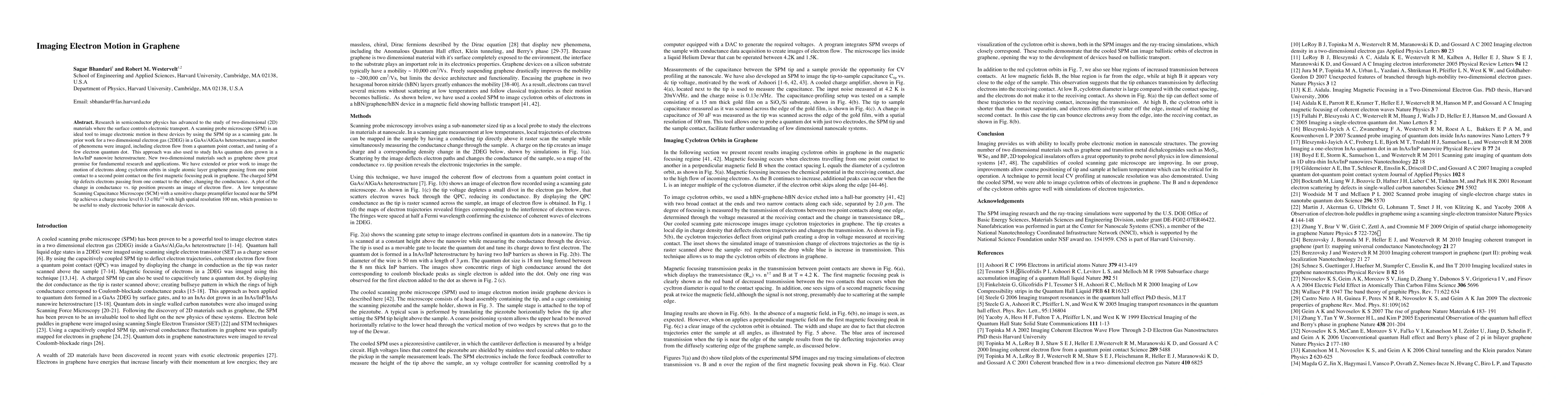
Research in semiconductor physics has advanced to the study of two-dimensional (2D) materials where the surface controls electronic transport. A scanning probe microscope (SPM) is an ideal tool to image electronic motion in these devices by using the SPM tip as a scanning gate. In prior work for a two dimensional electron gas (2DEG) in a GaAs/AlGaAs heterostructure, a number of phenomena were imaged, including electron flow from a quantum point contact, and tuning of a few electron quantum dot. This approach was also used to study InAs quantum dots grown in a InAs/InP nanowire heterostructure. New two-dimensional materials such as graphene show great promise for fundamental research and applications. We have extended or prior work to image the motion of electrons along cyclotron orbits in single atomic layer graphene passing from one point contact to a second point contact on the first magnetic focusing peak in graphene. The charged SPM tip defects electrons passing from one contact to the other, changing the conductance. A plot of the change in conductance vs. tip position presents an image of electron flow. A low temperature Scanning Capacitance Microscope (SCM) with a sensitive charge preamplifier located near the SPM tip achieves a charge noise level 0.13 e/Hz1/2 with high spatial resolution 100 nm, which promises to be useful to study electronic behavior in nanoscale devices.
AI Key Findings
Get AI-generated insights about this paper's methodology, results, significance, and more — seven facets brought into focus.
Impact
Paper Details
PDF Preview
Key Terms
Citation Network
Current paper (gray), citations (green), references (blue)
Display is limited for performance on very large graphs.
Discussion 0