Imaging ferroelectric domains with soft X-ray ptychography at the oxygen K-edge
Publication
Metrics
AI Quick Summary
This study visualizes ferroelectric domains in a BiFeO$_3$ film using ptychographic dichroic imaging with polarized soft X-rays at the O K-edge, revealing domain structure. The technique separates ferroelectric from antiferromagnetic contributions and can be applied to other multiferroic oxides.
Paper Preview
Abstract
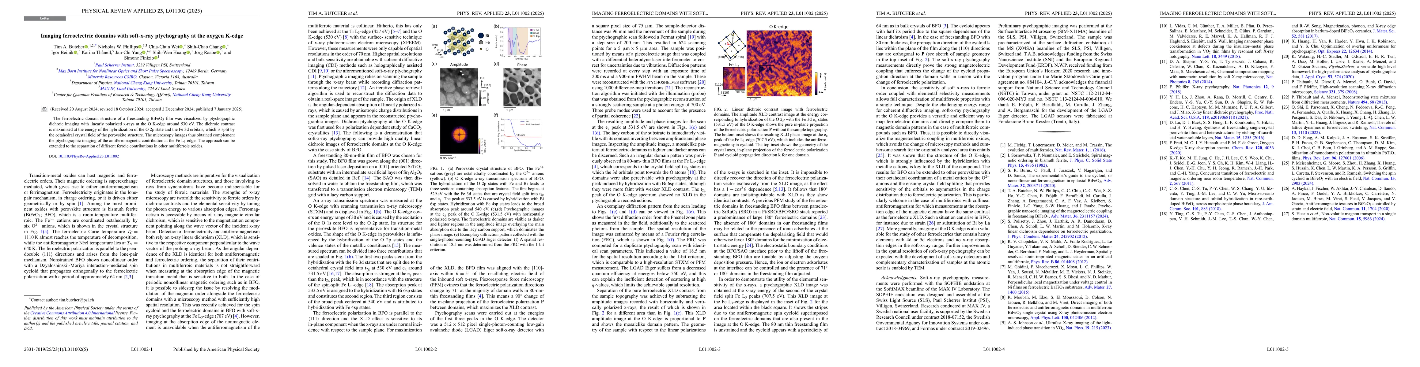
The ferroelectric domain structure of a freestanding BiFeO$_3$ film was visualized by ptychographic dichroic imaging with linearly polarized X-rays at the O K-edge around 530 eV. The dichroic contrast is maximized at the energy of the hybridization of the O 2p state and the Fe 3d orbitals, which is split by the octahedral crystal field of the perovskite structure. The thus obtained microscopy images compliment the ptychographic imaging of the antiferromagnetic contribution at the Fe L$_3$-edge. The approach is extendible to the separation of different ferroic contributions in other multiferroic oxides.
AI Key Findings
Get AI-generated insights about this paper's methodology, results, significance, and more — seven facets brought into focus.
Impact
Paper Details
Authors
PDF Preview
Citation Network
Current paper (gray), citations (green), references (blue)
Display is limited for performance on very large graphs.
Discussion 0