ArxivLens

Quick Summary:

This paper proposes using virtual mono-energetic reconstructions from spectral CT scanners to augment training data for myocardium segmentation in cardiac CT angiography (CCTA). Combining this with linear intensity scaling improves segmentation accuracy, achieving a Dice similarity coefficient of 0.901 $\pm$ 0.036 compared to 0.846 $\pm$ 0.125 with conventional images alone.

arXiv Id: 1810.03968
Date Published: 2019-01-29
Date Updated: 2019-01-29

Categories:

Key Terms (TF-IDF):

ct (0.287)
augmentation (0.285)
energetic (0.273)
virtual (0.236)
pm (0.209)
training data (0.197)
segmentation (0.193)
images (0.188)
data augmentation (0.184)
training (0.177)
acquired (0.173)
conventional (0.166)
agent (0.162)
contrast (0.158)
improves (0.144)
scaling (0.130)
spectral (0.115)
vs (0.110)
learning methods (0.105)
data (0.103)
License: http://arxiv.org/licenses/nonexclusive-distrib/1.0/

Summary:

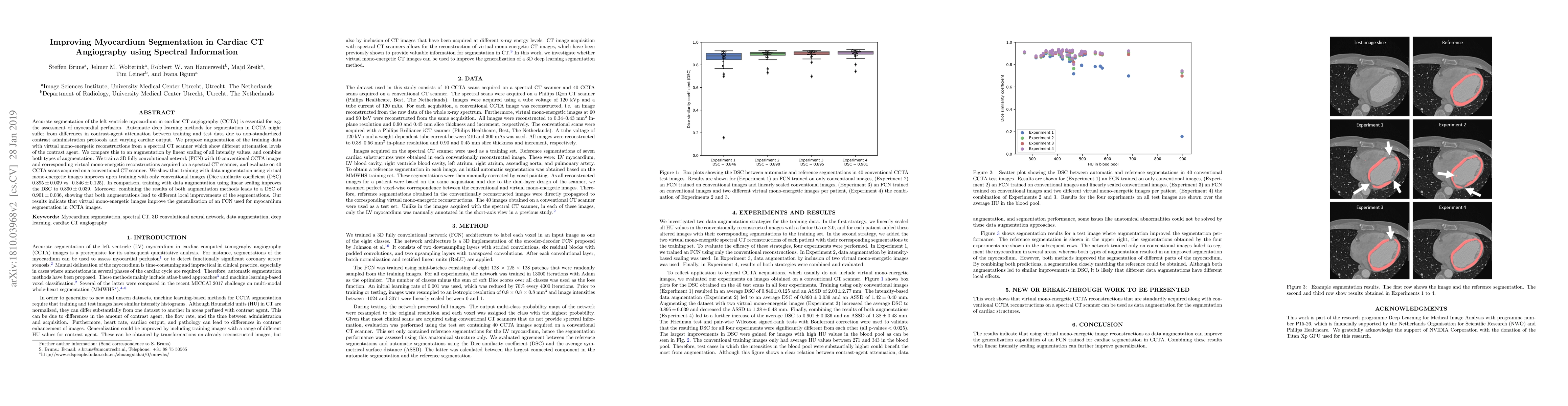
Accurate segmentation of the left ventricle myocardium in cardiac CT angiography (CCTA) is essential for e.g. the assessment of myocardial perfusion. Automatic deep learning methods for segmentation in CCTA might suffer from differences in contrast-agent attenuation between training and test data due to non-standardized contrast administration protocols and varying cardiac output. We propose augmentation of the training data with virtual mono-energetic reconstructions from a spectral CT scanner which show different attenuation levels of the contrast agent. We compare this to an augmentation by linear scaling of all intensity values, and combine both types of augmentation. We train a 3D fully convolutional network (FCN) with 10 conventional CCTA images and corresponding virtual mono-energetic reconstructions acquired on a spectral CT scanner, and evaluate on 40 CCTA scans acquired on a conventional CT scanner. We show that training with data augmentation using virtual mono-energetic images improves upon training with only conventional images (Dice similarity coefficient (DSC) 0.895 $\pm$ 0.039 vs. 0.846 $\pm$ 0.125). In comparison, training with data augmentation using linear scaling improves the DSC to 0.890 $\pm$ 0.039. Moreover, combining the results of both augmentation methods leads to a DSC of 0.901 $\pm$ 0.036, showing that both augmentations lead to different local improvements of the segmentations. Our results indicate that virtual mono-energetic images improve the generalization of an FCN used for myocardium segmentation in CCTA images.

Citation & Reference Network Visualization

This graph shows how this paper is connected to other research. The current paper is shown in dark gray, papers that cite it are in green, and papers it references are in blue. You can:

Current Paper
Papers citing this work (Citations)
Papers cited by this work (References)

• Hover over any node to see the paper title

• Click a node to view that paper's details

• Drag nodes to rearrange the graph

• Use the zoom controls (bottom right) to zoom in/out

Citations and References

Title Authors Year Actions
Deep learning-based whole-heart segmentation in 4D contrast-enhanced cardiac CT
Steffen Bruns, J. Wolterink, T. V. D. Boogert, J. Runge, B. Bouma, J. Henriques, J. Baan, M. Viergever, N. Planken, I. Išgum
2021
View Paper
A review of medical image data augmentation techniques for deep learning applications
P. Chlap, Hang Min, Nym Vandenberg, J. Dowling, L. Holloway, A. Haworth
2021
View Paper
Automatic whole-heart segmentation in 4D TAVI treatment planning CT
Steffen Bruns, J. Wolterink, T. V. D. Boogert, J. Henriques, J. Baan, Nils Planken, I. Išgum
2021
View Paper
CASPER: Conventional CT database Augmentation using deep learning based SPEctral CT images geneRation
P. Lartaud, Aymeric Rouchaud, R. Dessouky, A. Vlachomitrou, J. Rouet, O. Nempont, L. Boussel, P. Douek
2020
View Paper
Automatic quantification of myocardium and pericardial fat from coronary computed tomography angiography: a multicenter study
Xiuxiu He, B. Guo, Y. Lei, Tonghe Wang, W. Curran, Tian Liu, L. Zhang, Xiaofeng Yang
2020
View Paper
Deep Learning from Dual-Energy Information for Whole-Heart Segmentation in Dual-Energy and Single-Energy Non-Contrast-Enhanced Cardiac CT
Steffen Bruns, J. Wolterink, R. Takx, R. V. Hamersvelt, D. Suchá, M. Viergever, T. Leiner, I. Išgum
2020
View Paper
Artificial intelligence: improving the efficiency of cardiovascular imaging
A. Lin, Márton Kolossváry, I. Išgum, P. Maurovich-Horvát, P. Slomka, D. Dey
2020
View Paper
Automated Left Ventricular Myocardium Segmentation Using 3D Deeply Supervised Attention U-Net for Coronary Computed Tomography Angiography.
Bang Jun Guo, Xiuxiu He, Y. Lei, J. Harms, Tonghe Wang, W. Curran, Tian Liu, Long Jiang Zhang, Xiaofeng Yang
2020
View Paper

Comments (0)

This website uses cookies to ensure you get the best experience. Learn more