Summary

The field of optical microscopy spans across numerous industries and research domains, ranging from education to healthcare, quality inspection and analysis. Nonetheless, a key limitation often cited by optical microscopists refers to the limit of its lateral resolution (typically defined as ~200nm), with potential circumventions involving either costly external modules (e.g. confocal scan heads, etc) and/or specialized techniques [e.g. super-resolution (SR) fluorescent microscopy]. Addressing these challenges in a normal (non-specialist) context thus remains an aspect outside the scope of most microscope users & facilities. This study thus seeks to evaluate an alternative & economical approach to achieving SR optical microscopy, involving non-fluorescent phase-modulated microscopical modalities such as Zernike phase contrast (PCM) and differential interference contrast (DIC) microscopy. Two in silico deep neural network (DNN) architectures which we developed previously (termed O-Net and Theta-Net) are assessed on their abilities to resolve a custom-fabricated test target containing nanoscale features calibrated via atomic force microscopy (AFM). The results of our study demonstrate that although both O-Net and Theta-Net seemingly performed well when super-resolving these images, they were complementary (rather than competing) approaches to be considered for image SR, particularly under different image signal-to-noise ratios (SNRs). High image SNRs favoured the application of O-Net models, while low SNRs inclined preferentially towards Theta-Net models. These findings demonstrate the importance of model architectures (in conjunction with the source image SNR) on model performance and the SR quality of the generated images where DNN models are utilized for non-fluorescent optical nanoscopy, even where the same training dataset & number of epochs are being used.
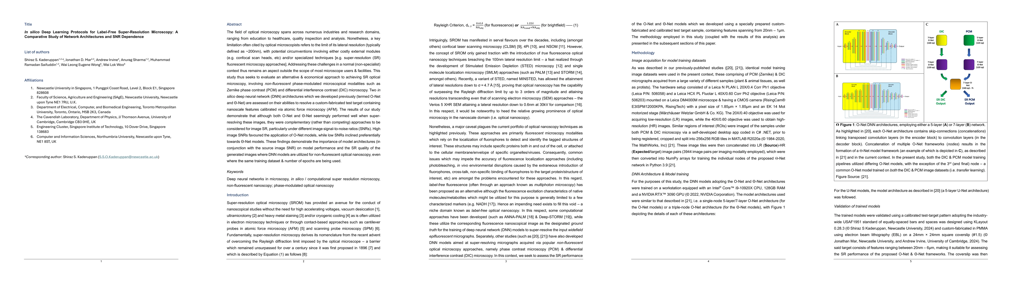
AI Key Findings

Generated Oct 01, 2025

Methodology

The study evaluated two deep neural network architectures, O-Net and Theta-Net, for super-resolution (SR) in label-free phase-modulated microscopy. They used a custom-fabricated test target with nanoscale features calibrated via atomic force microscopy (AFM) to assess image resolution under varying signal-to-noise ratios (SNRs).

Key Results

  • O-Net performed better at high SNR, while Theta-Net excelled under low SNR conditions.
  • Both models surpassed the Rayleigh diffraction limit of 308nm, resolving features down to 170nm (SR PCM) and 200nm (SR DIC).
  • Theta-Net generated more noise for features ≤200nm, whereas O-Net provided clearer delineation of nanoscale structures.

Significance

This research provides an economical alternative to traditional super-resolution techniques, enabling high-resolution imaging in non-specialist settings. It demonstrates the potential of deep learning for label-free nanoscopy, with implications for biomedical research, materials science, and quality control.

Technical Contribution

The study demonstrates how deep learning models can operate in the Fourier domain of the microscope's optical train to resolve features below the Rayleigh diffraction limit through learned convolution and deconvolution kernels.

Novelty

This work introduces a comparative analysis of O-Net and Theta-Net for label-free SR microscopy, showing their complementary performance based on SNR, and proposes a framework for integrating Fourier domain operations into DNN architectures for nanoscale imaging.

Limitations

  • The developed models are specific to the Leica DM4000M microscope and RisingCam® camera setup, requiring retraining for different optical configurations.
  • There is no objective metric for quantifying SR performance in non-fluorescent micrographs without ground truth images.

Future Work

  • Integrate the shear angle calculation from Nomarski prisms into the loss function to improve model resilience to pseudo-relief fluctuations.
  • Explore the use of Fourier Ring Correlation (FRC) and intensity profile analysis for objective resolution quantification in non-fluorescent micrographs.
  • Develop more complex DNN architectures with additional layers to enhance resolution capabilities.

Paper Details

PDF Preview

Download PDF

Comments (0)

This website uses cookies to ensure you get the best experience. Learn more