Publication
Metrics
AI Quick Summary
This study successfully achieved in vivo ultrasound-switchable fluorescence (USF) imaging in mice, demonstrating high-resolution imaging in porcine heart tissue and mouse breast tumors via local injections. The USF contrast agent proved stable in biological environments, primarily accumulating in the spleen, validated by micro-CT.
Paper Preview
Abstract
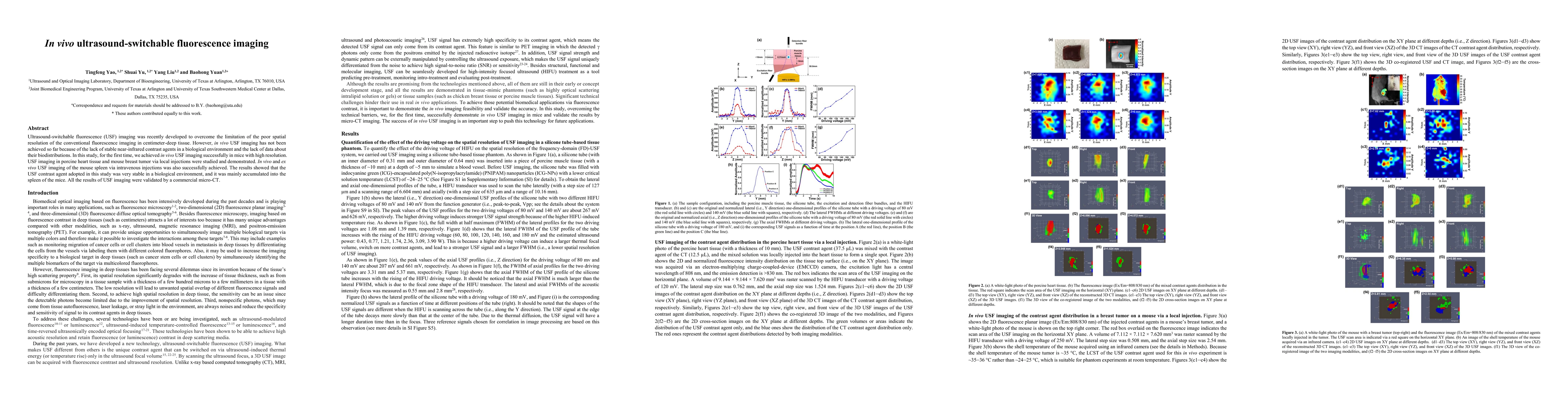
Ultrasound-switchable fluorescence (USF) imaging was recently developed to overcome the limitation of the poor spatial resolution of the conventional fluorescence imaging in centimeter-deep tissue. However, in vivo USF imaging has not been achieved so far because of the lack of stable near-infrared contrast agents in a biological environment and the lack of data about their biodistributions. In this study, for the first time, we achieved in vivo USF imaging successfully in mice with high resolution. USF imaging in porcine heart tissue and mouse breast tumor via local injections were studied and demonstrated. In vivo and ex vivo USF imaging of the mouse spleen via intravenous injections was also successfully achieved. The results showed that the USF contrast agent adopted in this study was very stable in a biological environment, and it was mainly accumulated into the spleen of the mice. All the results of USF imaging were validated by a commercial micro-CT.
AI Key Findings
Get AI-generated insights about this paper's methodology, results, significance, and more — seven facets brought into focus.
Impact
Paper Details
PDF Preview
Key Terms
Citation Network
Current paper (gray), citations (green), references (blue)
Display is limited for performance on very large graphs.
Discussion 0