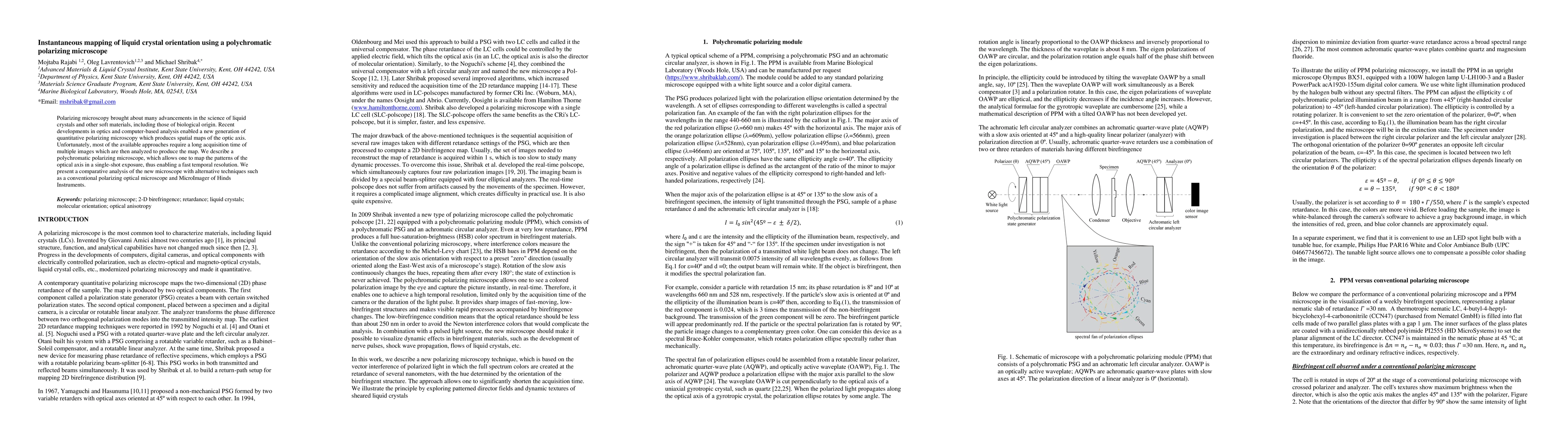
Instantaneous mapping of liquid crystal orientation using a polychromatic polarizing microscope
Publication
Metrics
AI Quick Summary
This paper introduces a polychromatic polarizing microscope that enables instantaneous mapping of liquid crystal orientation in a single-shot exposure, significantly improving temporal resolution compared to conventional methods. The study compares this new technique with traditional polarizing microscopy and MicroImager, highlighting its efficiency.
Paper Preview
Abstract
Polarizing microscopy brought about many advancements in the science of liquid crystals and other soft materials, including those of biological origin. Recent developments in optics and computer-based analysis enabled a new generation of quantitative polarizing microscopy which produces spatial maps of the optic axis. Unfortunately, most of the available approaches require a long acquisition time of multiple images which are then analyzed to produce the map. We describe a polychromatic polarizing microscope, which allows one to map the patterns of the optical axis in a single-shot exposure, thus enabling a fast temporal resolution. We present a comparative analysis of the new microscope with alternative techniques such as a conventional polarizing optical microscope and MicroImager of Hinds Instruments.
AI Key Findings
Get AI-generated insights about this paper's methodology, results, significance, and more — seven facets brought into focus.
Impact
Paper Details
Authors
PDF Preview
Key Terms
Citation Network
Current paper (gray), citations (green), references (blue)
Display is limited for performance on very large graphs.
Discussion 0