Researchers developed a new method for segmenting retinal layers from 3D optical coherence tomography images using diffusion maps, achieving robust results in low-contrast situations.
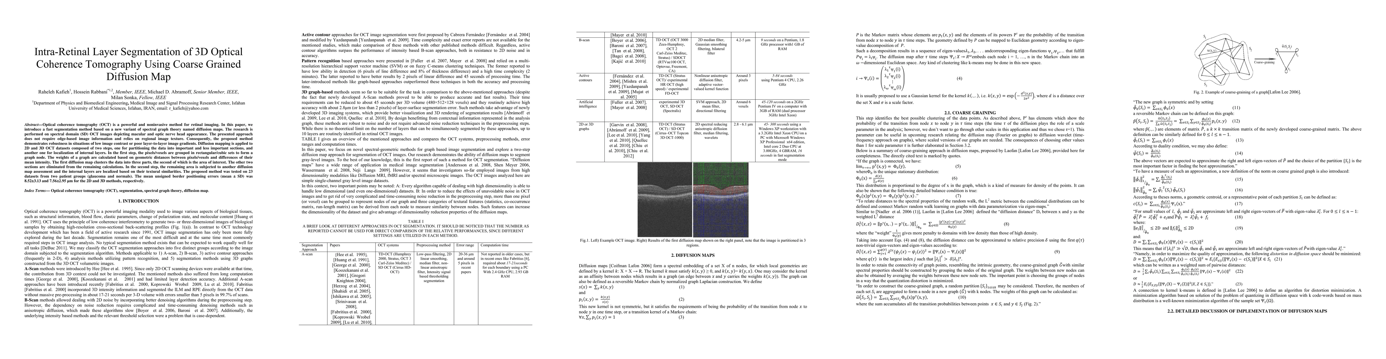
Optical coherence tomography (OCT) is a powerful and noninvasive method for retinal imaging. In this paper, we introduce a fast segmentation method based on a new variant of spectral graph theory named diffusion maps. The research is performed on spectral domain (SD) OCT images depicting macular and optic nerve head appearance. The presented approach does not require edge-based image information and relies on regional image texture. Consequently, the proposed method demonstrates robustness in situations of low image contrast or poor layer-to-layer image gradients. Diffusion mapping is applied to 2D and 3D OCT datasets composed of two steps, one for partitioning the data into important and less important sections, and another one for localization of internal layers.In the first step, the pixels/voxels are grouped in rectangular/cubic sets to form a graph node.The weights of a graph are calculated based on geometric distances between pixels/voxels and differences of their mean intensity.The first diffusion map clusters the data into three parts, the second of which is the area of interest. The other two sections are eliminated from the remaining calculations. In the second step, the remaining area is subjected to another diffusion map assessment and the internal layers are localized based on their textural similarities.The proposed method was tested on 23 datasets from two patient groups (glaucoma and normals). The mean unsigned border positioning errors(mean - SD) was 8.52 - 3.13 and 7.56 - 2.95 micrometer for the 2D and 3D methods, respectively.
Get AI-generated insights about this paper's methodology, results, and significance.
Current paper (gray), citations (green), references (blue)
Title | Authors | Year | Actions |
---|---|---|---|
Identifying the retinal layers from optical coherence tomography images using a 3D segmentation method
|
Akter Hossain, Andrea Giani, Victor Chong, S. Sivaprasad, Tasin R. Bhuiyan, Theodore Smith, Manaswini Pradhan, Alauddin Bhuiyan
|
2025
|
View Paper |
Enhancing OCT patch-based segmentation with improved GAN data augmentation and semi-supervised learning
|
J. Kugelman, D. Alonso-Caneiro, Scott A. Read, Stephen J. Vincent, Michael J. Collins
|
2024
|
View Paper |
SLO-Net: Enhancing Multiple Sclerosis Diagnosis Beyond Optical Coherence Tomography Using Infrared Reflectance Scanning Laser Ophthalmoscopy Images
|
R. Arian, A. Aghababaei, A. Soltanipour, Z. Khodabandeh, Sajed Rakhshani, Shwasa B Iyer, F. Ashtari, H. Rabbani, R. Kafieh
|
2024
|
View Paper |
Research of the Active Contour Method for Diagnostic Analysis of Regions of Interest in Optical Coherence Tomography Images of Retinal Layers
|
N. Ilyasova, Dmitry Doruzhinsky, N. Demin
|
2024
|
View Paper |
Boundary-Repairing Dual-Path Network for Retinal Layer Segmentation in OCT Image with Pigment Epithelial Detachment.
|
Xiaoming Liu, Xiao Li, Ying Zhang, Man Wang, Junping Yao, Jinshan Tang
|
2024
|
View Paper |
An efficient clustering algorithm for image segmentation of retinal OCT images
|
M. Bangar, Prachi Chaudhary
|
2024
|
View Paper |
Tensor Ring Decomposition Guided Dictionary Learning for OCT Image Denoising
|
P. G. Daneshmand, Hossein Rabbani
|
2024
|
View Paper |
Retinal OCT Image Denoising Based on Adaptive Bessel K-form Modeling
|
Sahar Jorjandi, Z. Amini, Maryam Samieinasab, Hossein Rabbani
|
2023
|
View Paper |
Retinal Layer Segmentation in OCT Images With Boundary Regression and Feature Polarization
|
Yubo Tan, W. Shen, Ming-Yuan Wu, Gui-Na Liu, Shixuan Zhao, Yang Chen, Kai-Fu Yang, Yongjie Li
|
2023
|
View Paper |
Discrimination of multiple sclerosis using OCT images from two different centers.
|
Z. Khodabandeh, H. Rabbani, F. Ashtari, H. Zimmermann, S. Motamedi, A. Brandt, Friedemann Paul, R. Kafieh
|
2023
|
View Paper |
A bibliometric analysis of artificial intelligence applications in macular edema: exploring research hotspots and Frontiers
|
Haiwen Feng, Jiaqi Chen, Zhichang Zhang, Yanfeng Lou, Shaochong Zhang, Weihua Yang
|
2023
|
View Paper |
Computational Intelligent Techniques and Analysis on Automated Retinal Layer Segmentation and Detection of Retinal Diseases
|
T. M. Sheeba, Applications, S. Albert, Antony Raj, M. Anand
|
2023
|
View Paper |
Clinical Applications of Artificial Intelligence in Glaucoma
|
Siamak Yousefi
|
2023
|
View Paper |
Automated assessment of the smoothness of retinal layers in optical coherence tomography images using a machine learning algorithm
|
J. Saeidian, Tahereh Mahmoudi, H. Riazi-Esfahani, Zahra Montazeriani, A. Khodabande, M. Zarei, Nazanin Ebrahimiadib, Behzad Jafari, A. Aghaei, Hossein Azimi, E. K. Pour
|
2023
|
View Paper |
Robust multi-view approaches for retinal layer segmentation in glaucoma patients via transfer learning
|
Mateo Gende, J. de Moura, J. Fernández-Vigo, J. Martínez-de-la-Casa, J. García-Feijóo, J. Novo, M. Ortega
|
2023
|
View Paper |
Human Coronary Plaque Optical Coherence Tomography Image Repairing, Multilayer Segmentation and Impact on Plaque Stress/Strain Calculations
|
Mengde Huang, A. Maehara, D. Tang, Jian Zhu, Liang Wang, R. Lv, Yanwen Zhu, Xiaoguo Zhang, M. Matsumura, Lijuan Chen, G. Ma, G. Mintz
|
2022
|
View Paper |
LOCTseg: A lightweight fully convolutional network for end-to-end optical coherence tomography segmentation
|
Esther Parra-Mora, L. Cruz
|
2022
|
View Paper |
Synthetic OCT Data Generation to Enhance the Performance of Diagnostic Models for Neurodegenerative Diseases
|
H. Danesh, D. Steel, J. Hogg, F. Ashtari, W. Innes, J. Bacardit, A. Hurlbert, J. Read, R. Kafieh
|
2022
|
View Paper |
A comparison of deep learning U-Net architectures for posterior segment OCT retinal layer segmentation
|
J. Kugelman, Joseph Allman, Scott A. Read, Stephen J. Vincent, Janelle Tong, M. Kalloniatis, F. Chen, M. Collins, D. Alonso-Caneiro
|
2022
|
View Paper |
Outer retinal thickness and visibility of the choriocapillaris in four distinct retinal regions imaged with spectral domain optical coherence tomography in dogs and cats
|
Elisa Mischi, Petr Soukup, Christine D Harman, Kazuya Oikawa, M. Kowalska, S. Hartnack, G. McLellan, A. Komáromy, S. Pot
|
2022
|
View Paper |
JIEB-linknet
|
M. N. Meeral, S. Shajun, M. N. Meeral, Research Scholar, M. M. Sathik
|
2022
|
View Paper |
Challenges for ocular disease identification in the era of artificial intelligence
|
Neha Gour, M. Tanveer, P. Khanna
|
2022
|
View Paper |
Automatic Segmentation of the Retinal Nerve Fiber Layer by Means of Mathematical Morphology and Deformable Models in 2D Optical Coherence Tomography Imaging
|
R. Berenguer-Vidal, R. Verdú, J. Morales-Sánchez, Inmaculada Sellés-Navarro, Rocío del Amor, Gabriel García, V. Naranjo
|
2021
|
View Paper |
Synthetic OCT data in challenging conditions: three-dimensional OCT and presence of abnormalities
|
H. Danesh, K. Maghooli, A. Dehghani, R. Kafieh
|
2021
|
View Paper |
Statistical modeling of retinal optical coherence tomography using the Weibull mixture model.
|
Sahar Jorjandi, Z. Amini, G. Plonka, H. Rabbani
|
2021
|
View Paper |
Automated retinal boundary segmentation of optical coherence tomography images using an improved Canny operator
|
Jian Liu, S. Yan, N. Lu, Dongni Yang, Hongyu Lv, Shuanglian Wang, Xin Zhu, Yuqian Zhao, Yi Wang, Zhenhe Ma, Yao Yu
|
2021
|
View Paper |
Retinal OCT Texture Analysis for Differentiating Healthy Controls from Multiple Sclerosis (MS) with/without Optic Neuritis
|
Hamidreza Dehghan Tazarjani, Z. Amini, R. Kafieh, F. Ashtari, E. Sadeghi
|
2021
|
View Paper |
Assessing the validity of a cross-platform retinal image segmentation tool in normal and diseased retina
|
Varsha Alex, T. Motevasseli, W. Freeman, J. Jayamon, D. Bartsch, S. Borooah
|
2021
|
View Paper |
Artificial intelligence in OCT angiography
|
T. Hormel, T. Hwang, Steven T. Bailey, David J. Wilson, David Huang, Yali Jia
|
2021
|
View Paper |
Livelayer: a semi-automatic software program for segmentation of layers and diabetic macular edema in optical coherence tomography images
|
Mansooreh Montazerin, Zahra Sajjadifar, Elias Khalili pour, H. Riazi-Esfahani, Tahereh Mahmoudi, H. Rabbani, Hossein Movahedian, A. Dehghani, M. Akhlaghi, R. Kafieh
|
2021
|
View Paper |
Diagnosis of Age Related Macular Degeneration by Curve Fitting RPE Layer
|
D. Pavithra, R. Vanithamani, E. Karolinekersin
|
2021
|
View Paper |
Evaluation of Asymmetry in Right and Left Eyes of Normal Individuals Using Extracted Features from Optical Coherence Tomography and Fundus Images
|
Tahereh Mahmudi, R. Kafieh, H. Rabbani, Alireza Mehri, M. Akhlaghi
|
2021
|
View Paper |
Optical Coherence Tomography in Neuromyelitis Optica spectrum disorder and Multiple Sclerosis: A population-based study.
|
F. Ashtari, Akram Ataei, R. Kafieh, Z. Khodabandeh, Mahdi Barzegar, Marzieh Raei, A. Dehghani, M. Mansurian
|
2020
|
View Paper |
A Multi-task Framework for Topology-guaranteed Retinal Layer Segmentation in OCT Images
|
Jun Cao, Xiaoming Liu, Ying Zhang, Man Wang
|
2020
|
View Paper |
Mathematical analysis of texture indicators for the segmentation of optical coherence tomography images
|
M. Monemian, H. Rabbani
|
2020
|
View Paper |
Assignment Flow for Order-Constrained OCT Segmentation
|
Dmitrij Sitenko, Bastian Boll, C. Schnörr
|
2020
|
View Paper |
Automatic Production of Synthetic Labeled OCT Images Using Active Shape Model
|
H. Danesh, K. Maghooli, R. Kafieh, A. Dehghani
|
2020
|
View Paper |
Deep learning architectures analysis for age-related macular degeneration segmentation on optical coherence tomography scans
|
K. Alsaih, M. Yusoff, T. Tang, I. Faye, F. Mériaudeau
|
2020
|
View Paper |
Forming Optimal Projection Images from Intra-Retinal Layers Using Curvelet-Based Image Fusion Method
|
Jalil Jalili, H. Rabbani, A. M. Dehnavi, R. Kafieh, M. Akhlaghi
|
2020
|
View Paper |
Towards Label-Free 3D Segmentation of Optical Coherence Tomography Images of the Optic Nerve Head Using Deep Learning
|
S. Devalla, T. Pham, S. Panda, Zhang Liang, Giridhar Subramanian, Anirudh Swaminathan, Chin Zhi Yun, M. Rajan, S. Mohan, R. Krishnadas, Vijay S Senthil, J. Leon, T. A. Tun, Ching-Yu Cheng, L. Schmetterer, S. Perera, T. Aung, Alexandre Hoang Thiery, M. Girard
|
2020
|
View Paper |
Automated Deformation-Based Analysis of 3D Optical Coherence Tomography in Diabetic Retinopathy
|
Maziyar M. Khansari, Jiong Zhang, Y. Qiao, J. Gahm, Mona Sharifi Sarabi, A. Kashani, Yonggang Shi
|
2020
|
View Paper |
Visualising ganglion cell layer based on image entropy optimisation for adaptive contrast enhancement
|
J.‐H. Han, J. Cha
|
2020
|
View Paper |
Fluid Segmentation in Neutrosophic Domain
|
Elyas Rashno, A. Rashno, S. Fadaei
|
2019
|
View Paper |
Local comparison of cup to disc ratio in right and left eyes based on fusion of color fundus images and OCT B-scans
|
Marzieh Mokhtari, H. Rabbani, A. M. Dehnavi, R. Kafieh, M. Akhlaghi, M. Pourazizi, Leyuan Fang
|
2019
|
View Paper |
Automated Layer Segmentation of Retinal Optical Coherence Tomography Images Using a Deep Feature Enhanced Structured Random Forests Classifier
|
Xiaoming Liu, Tianyu Fu, Zhifang Pan, Dong Liu, Wei Hu, Jun Liu, Kai Zhang
|
2019
|
View Paper |
Automatic Multifaceted Matlab Package for Analysis of Ocular Images (AMPAO)
|
R. Kafieh, Z. Amini, H. Rabbani, Bahareh Kaviani Baghbaderani, Bahareh Salafian, Fatemeh Mazaheri, Marzieh Mokhtari
|
2019
|
View Paper |
Level Set Method in Medical Imaging Segmentation
|
A. El-Baz, J. Suri
|
2019
|
View Paper |
Segmentation of mouse skin layers in optical coherence tomography image data using deep convolutional neural networks.
|
Timo Kepp, C. Droigk, M. Casper, M. Evers, G. Hüttmann, Nunciada Salma, D. Manstein, M. Heinrich, H. Handels
|
2019
|
View Paper |
A Mechanical Approach for Smooth Surface Fitting to Delineate Vessel Walls in Optical Coherence Tomography Images
|
Max L. Olender, L. Athanasiou, J. M. de la Torre Hernandez, Eyal Ben-Assa, F. R. Nezami, E. Edelman
|
2019
|
View Paper |
Detection of retinal pigment epithelium detachment from OCT images using multiscale Gaussian filtering
|
Meletios Liaskos, P. Asvestas, G. Matsopoulos, Alexander C Charonis, V. Anastassopoulos
|
2019
|
View Paper |
Topology-Preserving Shape-Based Regression Of Retinal Layers In Oct Image Data Using Convolutional Neural Networks
|
Timo Kepp, J. Ehrhardt, M. Heinrich, G. Hüttmann, H. Handels
|
2019
|
View Paper |
Automatic retinal layer segmentation in SD-OCT images with CSC guided by spatial characteristics
|
Kun Gao, W. Kong, Sijie Niu, Dengwang Li, Yuehui Chen
|
2019
|
View Paper |
Automatic Choroid Layer Segmentation from Optical Coherence Tomography Images Using Deep Learning
|
Saleha Masood, R. Fang, P. Li, Huating Li, Bin Sheng, Akash Mathavan, Xiangning Wang, Po Yang, Qiang Wu, J. Qin, W. Jia
|
2019
|
View Paper |
Robust segmentation of retinal layers in optical coherence tomography images based on a multistage active contour model
|
A. González-López, J. de Moura, J. Novo, M. Ortega, M. Penedo
|
2019
|
View Paper |
Automatic Retinal Layer Segmentation of OCT Images With Central Serous Retinopathy
|
Dehui Xiang, Geng Chen, Fei Shi, Weifang Zhu, Qinghuai Liu, Songtao Yuan, Xinjian Chen
|
2019
|
View Paper |
Multi-Modal Medical Image Registration with Full or Partial Data: A Manifold Learning Approach
|
Fereshteh S. Bashiri, Ahmadreza Baghaie, Reihaneh Rostami, Zeyun Yu, R. D'Souza
|
2018
|
View Paper |
Variational and PDE-based methods for image processing
|
J. Duan
|
2018
|
View Paper |
Three-dimensional texture analysis of optical coherence tomography images of ovarian tissue
|
T. Sawyer, Swati Chandra, Photini F S Rice, Jennifer W. Koevary, J. Barton
|
2018
|
View Paper |
Automatic segmentation of OCT retinal boundaries using recurrent neural networks and graph search.
|
J. Kugelman, D. Alonso-Caneiro, Scott A. Read, Stephen J. Vincent, M. Collins
|
2018
|
View Paper |
Intra-Retinal Layer Segmentation of Optical Coherence Tomography Using 3D Fully Convolutional Networks
|
Farkhondeh Kiaee, H. Fahimi, H. Rabbani
|
2018
|
View Paper |
Early diabetic retinopathy diagnosis based on local retinal blood vessel analysis in optical coherence tomography angiography (OCTA) images
|
Nabila Eladawi, M. Elmogy, F. Khalifa, M. Ghazal, N. Ghazi, A. Aboelfetouh, A. Riad, H. Sandhu, S. Schaal, A. El-Baz
|
2018
|
View Paper |
Functional imaging of ganglion and receptor cells in living human retina by osmotic contrast
|
Clara Pfaffle, Dierck Hillmann, H. Spahr, Lisa Kutzner, Sazan Burhan, Felix Hilge, Y. Miura, G. Huttmann
|
2018
|
View Paper |
Robust layer segmentation of esophageal OCT images based on graph search using edge-enhanced weights
|
Meng Gan, Cong Wang, Ting Yang, Na Yang, Miao Zhang, Wu Yuan, Xingde Li, Lirong Wang
|
2018
|
View Paper |
Automatic Segmentation of Retinal Layer in OCT Images With Choroidal Neovascularization
|
Dehui Xiang, Haihong Tian, Xiaoling Yang, Fei Shi, Weifang Zhu, H. Chen, Xinjian Chen
|
2018
|
View Paper |
OCT Fluid Segmentation using Graph Shortest Path and Convolutional Neural Network*
|
A. Rashno, D. Koozekanani, K. Parhi
|
2018
|
View Paper |
An approach to automated retinal layer segmentation in SDOCT images
|
M. G, Hari Krishnan G, H. R. J.
|
2018
|
View Paper |
Automatic Evaluation and Predictive Analysis of Optic Nerve Head for the Detection of Glaucoma
|
Kaushik Dutta, Rishav Mukherjee, Shamik Kundu, Tanmay Biswas, Anindya Sen
|
2018
|
View Paper |
Fully Automated Segmentation of Fluid/Cyst Regions in Optical Coherence Tomography Images With Diabetic Macular Edema Using Neutrosophic Sets and Graph Algorithms
|
A. Rashno, D. Koozekanani, P. Drayna, Behzad Nazari, S. Sadri, H. Rabbani, K. Parhi
|
2018
|
View Paper |
Three Dimensional Fully Convolutional Networks for Segmentation of Optical Coherence Tomography Images in Neurodegenerative Disease
|
Farkhondeh Kiaee, H. Fahimi, R. Kafieh, A. Brandt, H. Rabbani
|
2018
|
View Paper |
An integrated framework for automatic clinical assessment of diabetic retinopathy grade using spectral domain OCT images
|
Ahmed ElTanboly, M. Ghazal, A. Khalil, A. Shalaby, Ali M. Mahmoud, A. Switala, M. El-Azab, S. Schaal, A. El-Baz
|
2018
|
View Paper |
Macular OCT Classification Using a Multi-Scale Convolutional Neural Network Ensemble
|
Reza Rasti, H. Rabbani, A. Mehridehnavi, Fedra Hajizadeh
|
2018
|
View Paper |
Retracted: Early diabetic retinopathy diagnosis based on local retinal blood vessels analysis in optical coherence tomography angiography (OCTA) images.
|
Nabila Eladawi, Mohammed M Elmogy, F. Khalifa, M. Ghazal, N. Ghazi, A. Aboelfetouh, A. Riad, H. Sandhu, S. Schaal, A. El-Baz
|
2018
|
View Paper |
Deep learning approach for the detection and quantification of intraretinal cystoid fluid in multivendor optical coherence tomography.
|
F. Venhuizen, B. van Ginneken, B. Liefers, F. van Asten, V. Schreur, S. Fauser, C. Hoyng, T. Theelen, C. I. Sánchez
|
2018
|
View Paper |
Superpixel guided active contour segmentation of retinal layers in OCT volumes
|
Fangliang Bai, S. Gibson, M. Marques, A. Podoleanu
|
2018
|
View Paper |
Automatic diagnosis of abnormal macula in retinal optical coherence tomography images using wavelet-based convolutional neural network features and random forests classifier
|
Reza Rasti, A. Mehridehnavi, H. Rabbani, Fedra Hajizadeh
|
2018
|
View Paper |
Machine learning and data science in soft materials engineering
|
Andrew L. Ferguson
|
2018
|
View Paper |
A hybrid method for 3D mosaicing of OCT images of macula and Optic Nerve Head
|
Alieh Ahdi, H. Rabbani, Alireza Vard
|
2017
|
View Paper |
Wavelet-based Convolutional Mixture of Experts model: An application to automatic diagnosis of abnormal macula in retinal optical coherence tomography images
|
Reza Rasti, A. Mehridehnavi, H. Rabbani, Fedra Hajizadeh
|
2017
|
View Paper |
Fully-automated segmentation of fluid regions in exudative age-related macular degeneration subjects: Kernel graph cut in neutrosophic domain
|
A. Rashno, Behzad Nazari, D. Koozekanani, P. Drayna, S. Sadri, H. Rabbani, K. Parhi
|
2017
|
View Paper |
Cystoid macular edema segmentation of Optical Coherence Tomography images using fully convolutional neural networks and fully connected CRFs
|
Fangliang Bai, M. Marques, S. Gibson
|
2017
|
View Paper |
Optical coherence tomography image denoising using Gaussianization transform.
|
Z. Amini, H. Rabbani
|
2017
|
View Paper |
A deep convolutional feature based learning layer-specific edges method for segmenting OCT image
|
Tianyu Fu, Xiaoming Liu, Dong Liu, Zhou Yang
|
2017
|
View Paper |
Statistical modeling of Optical Coherence Tomography images by asymmetric Normal Laplace mixture model
|
Sahar Jorjandi, H. Rabbani, R. Kafieh, Z. Amini
|
2017
|
View Paper |
Robust total retina thickness segmentation in optical coherence tomography images using convolutional neural networks.
|
F. Venhuizen, B. van Ginneken, B. Liefers, Mark J. J. P. van Grinsven, S. Fauser, C. Hoyng, T. Theelen, C. I. Sánchez
|
2017
|
View Paper |
Automatic segmentation of nine retinal layer boundaries in OCT images of non-exudative AMD patients using deep learning and graph search.
|
Leyuan Fang, David Cunefare, Chong Wang, R. Guymer, Shutao Li, Sina Farsiu
|
2017
|
View Paper |
ReLayNet: Retinal Layer and Fluid Segmentation of Macular Optical Coherence Tomography using Fully Convolutional Network
|
Abhijit Guha Roy, Sailesh Conjeti, S. Karri, Debdoot Sheet, A. Katouzian, C. Wachinger, Nassir Navab
|
2017
|
View Paper |
Involuntary eye motion correction in retinal optical coherence tomography: Hardware or software solution?
|
Ahmadreza Baghaie, Zeyun Yu, R. D'Souza
|
2017
|
View Paper |
Automated intraretinal segmentation of SD-OCT images in normal and age-related macular degeneration eyes.
|
L. de Sisternes, G. Jonna, Jason Moss, M. Marmor, T. Leng, D. Rubin
|
2017
|
View Paper |
A computer‐aided diagnostic system for detecting diabetic retinopathy in optical coherence tomography images
|
Ahmed ElTanboly, M. Ismail, A. Shalaby, A. Switala, A. El-Baz, S. Schaal, G. Gimel'farb, M. El-Azab
|
2017
|
View Paper |
Segmentation of Locally Varying Numbers of Outer Retinal Layers by a Model Selection Approach
|
J. Novosel, S. Yzer, Koen A. Vermeer, L. V. van Vliet
|
2017
|
View Paper |
Joint Segmentation of Retinal Layers and Focal Lesions in 3-D OCT Data of Topologically Disrupted Retinas
|
J. Novosel, Koen A. Vermeer, Jan H. de Jong, Ziyuan Wang, L. V. van Vliet
|
2017
|
View Paper |
Individual Drusen Segmentation and Repeatability and Reproducibility of Their Automated Quantification in Optical Coherence Tomography Images
|
L. de Sisternes, G. Jonna, Margaret A. Greven, Qiang Chen, T. Leng, D. Rubin
|
2017
|
View Paper |
Transfer learning based classification of optical coherence tomography images with diabetic macular edema and dry age-related macular degeneration.
|
S. Karri, Debjani Chakraborty, J. Chatterjee
|
2017
|
View Paper |
Classifying DME vs normal SD-OCT volumes: A review
|
J. Massich, M. Rastgoo, G. Lemaître, C. Cheung, T. Wong, D. Sidibé, F. Mériaudeau
|
2016
|
View Paper |
Automatic choroid layer segmentation: A slicing approach
|
Chengyu Luo
|
2016
|
View Paper |
Intra-retinal layers segmentation of macular OCT images based on the graph optimal approach
|
Zhijun Gao, Wei Bu, Xiangqian Wu, Yalin Zheng
|
2016
|
View Paper |
Sector mapping method for 3D detached retina visualization
|
Yi-ran Zhai, Yong Zhao, Jie Zhong, Ke Li, Cui-xin Lu, Bing Zhang
|
2016
|
View Paper |
Automated segmentation of retinal layers from optical coherence tomography images using geodesic distance
|
J. Duan, C. Tench, I. Gottlob, F. Proudlock, L. Bai
|
2016
|
View Paper |
Retinal layer delineation through learning of tissue photon interaction in optical coherence tomography
|
S. Karri, Niladri Garai, Debaleena Nawn, Sambuddha Ghosh, Debjani Chakraborty, J. Chatterjee
|
2016
|
View Paper |
Alignment of optic nerve head optical coherence tomography B-scans in right and left eyes
|
Marzieh Mokhtari, H. Rabbani, Alireza Mehri-Dehnavi
|
2016
|
View Paper |
Retinal automatic segmentation method based on prior information and optimized boundary tracking algorithm
|
Dongmei Fu, Hejun Tong, Ling Luo, Fulin Gao
|
2016
|
View Paper |
Optical Coherence Tomography Image Analysis
|
H. Rabbani, R. Kafieh, Z. Amini
|
2016
|
View Paper |
3D reconstruction of cochlea using optical coherence tomography
|
Tuukka Karvonen, Yuuki Uranishi, T. Sakamoto, Yosuke Tona, K. Okamoto, H. Tamura, T. Kuroda
|
2016
|
View Paper |
Classification of SD-OCT Volumes Using Local Binary Patterns: Experimental Validation for DME Detection
|
G. Lemaître, M. Rastgoo, J. Massich, C. Cheung, T. Wong, E. Lamoureux, D. Milea, F. Mériaudeau, D. Sidibé
|
2016
|
View Paper |
Learning layer-specific edges for segmenting retinal layers with large deformations.
|
S. Karri, Debjani Chakraborthi, J. Chatterjee
|
2016
|
View Paper |
An automated framework for 3D serous pigment epithelium detachment segmentation in SD-OCT images
|
Z. Sun, H. Chen, Fei Shi, Lirong Wang, Weifang Zhu, Dehui Xiang, Chenglin Yan, Liang Li, Xinjian Chen
|
2016
|
View Paper |
Statistical Modeling of Retinal Optical Coherence Tomography
|
Z. Amini, H. Rabbani
|
2016
|
View Paper |
Loosely coupled level sets for simultaneous 3D retinal layer segmentation in optical coherence tomography
|
J. Novosel, G. Thepass, H. Lemij, J. Boer, Koen A. Vermeer, L. Vliet
|
2015
|
View Paper |
A web-based framework for anatomical assessment of the retina using OCT
|
A. González-López, M. Ortega, M. Penedo, Pablo Charlón
|
2015
|
View Paper |
Classification of SD-OCT Volumes with LBP: Application to DME Detection
|
G. Lemaître, M. Rastgoo, J. Massich, Shrinivasan Sankar, F. Mériaudeau, D. Sidibé
|
2015
|
View Paper |
Automated Layer Segmentation of 3D Macular Images Using Hybrid Methods
|
Chuang Wang, Yaxing Wang, Djibril Kaba, Zidong Wang, Xiaohui Liu, Yongmin Li
|
2015
|
View Paper |
Segmentation of Intra-retinal Layers in 3D Optic Nerve Head Images
|
Chuang Wang, Yaxing Wang, Djibril Kaba, Haogang Zhu, Y. Lv, Zidong Wang, Xiaohui Liu, Yongmin Li
|
2015
|
View Paper |
3D Automatic Segmentation Method for Retinal Optical Coherence Tomography Volume Data Using Boundary Surface Enhancement
|
Yankui Sun, Tian Zhang, Yue Zhao, Yufan He
|
2015
|
View Paper |
RefMoB, a Reflectivity Feature Model-Based Automated Method for Measuring Four Outer Retinal Hyperreflective Bands in Optical Coherence Tomography.
|
Douglas Ross, Mark E. Clark, P. Godara, C. Huisingh, G. McGwin, C. Owsley, K. M. Litts, R. Spaide, K. Sloan, C. Curcio
|
2015
|
View Paper |
Visual Prognosis of Eyes Recovering From Macular Hole Surgery Through Automated Quantitative Analysis of Spectral-Domain Optical Coherence Tomography (SD-OCT) Scans.
|
L. de Sisternes, Julia Hu, D. Rubin, T. Leng
|
2015
|
View Paper |
Three Dimensional Data-Driven Multi Scale Atomic Representation of Optical Coherence Tomography
|
R. Kafieh, H. Rabbani, I. Selesnick
|
2015
|
View Paper |
Thickness Mapping of Eleven Retinal Layers Segmented Using the Diffusion Maps Method in Normal Eyes
|
R. Kafieh, H. Rabbani, Fedra Hajizadeh, M. Abràmoff, M. Sonka
|
2015
|
View Paper |
Automated age-related macular degeneration classification in OCT using unsupervised feature learning
|
F. Venhuizen, B. Ginneken, Bart Bloemen, M. V. Grinsven, R. Philipsen, C. Hoyng, T. Theelen, C. I. Sánchez
|
2015
|
View Paper |
Automated pharmaceutical tablet coating layer evaluation of optical coherence tomography images
|
D. Markl, G. Hannesschläger, S. Sacher, M. Leitner, J. Khinast, A. Buchsbaum
|
2015
|
View Paper |
Automated 3-D Retinal Layer Segmentation of Macular Optical Coherence Tomography Images With Serous Pigment Epithelial Detachments
|
Fei Shi, Xinjian Chen, Heming Zhao, Weifang Zhu, Dehui Xiang, Enting Gao, M. Sonka, Haoyu Chen
|
2015
|
View Paper |
Three-dimensional image classification using hierarchical spatial decomposition : a study using retinal data
|
A. Albarrak
|
2015
|
View Paper |
A comparative study on macular optical coherence tomography image alignment
|
Annan Li, Jun Cheng, D. Wong, Jiang Liu
|
2014
|
View Paper |
Structure-preserving multi-scale decompositions for skin and retinal OCT image
|
Annan Li, Jun Cheng, D. Wong, H. Tey, Jiang Liu
|
2014
|
View Paper |
State-of-the-Art in Retinal Optical Coherence Tomography Image Analysis
|
Ahmadreza Baghaie, R. D'Souza, Zeyun Yu
|
2014
|
View Paper |
Automated retinal layers segmentation in SD-OCT images using dual-gradient and spatial correlation smoothness constraint
|
Sijie Niu, Qiang Chen, L. D. Sisternes, D. Rubin, Weiwei Zhang, Qinghuai Liu
|
2014
|
View Paper |
An Automatic Algorithm for Segmentation of the Boundaries of Corneal Layers in Optical Coherence Tomography Images using Gaussian Mixture Model
|
Mahdi Kazemian Jahromi, R. Kafieh, H. Rabbani, A. M. Dehnavi, A. Peyman, Fedra Hajizadeh, Mohammadreza Ommani
|
2014
|
View Paper |
User-guided segmentation for volumetric retinal optical coherence tomography images.
|
Xin Yin, J. Chao, Ruikang K. Wang
|
2014
|
View Paper |
Multi-Surface and Multi-Field Co-Segmentation of 3-D Retinal Optical Coherence Tomography
|
H. Bogunović, M. Sonka, Young H. Kwon, P. Kemp, M. Abràmoff, Xiaodong Wu
|
2014
|
View Paper |
Combination of graph theoretic grouping and time-frequency analysis for image segmentation
|
R. Kafieh, H. Rabbani, S. Gazor
|
2014
|
View Paper |
A statistical model for 3D segmentation of retinal choroid in optical coherence tomography images
|
Fahimeh Ghasemi, Hossein Rabbani
|
2014
|
View Paper |
Comparison of macular OCTs in right and left eyes of normal people
|
Tahereh Mahmudi, R. Kafieh, H. Rabbani, A. Mehri dehnavi, Mohammadreza Akhlagi
|
2014
|
View Paper |
Thickness Mapping of Eleven Retinal Layers in Normal Eyes Using Spectral Domain Optical Coherence Tomography
|
R. Kafieh, H. Rabbani, Fedra Hajizadeh, M. Abràmoff, M. Sonka
|
2013
|
View Paper |
Vessel segmentation in images of optical coherence tomography using shadow information and thickening of Retinal Nerve Fiber Layer
|
R. Kafieh, H. Danesh, H. Rabbani, M. Abràmoff, M. Sonka
|
2013
|
View Paper |
Forming projection images from each layer of retina using diffusion may based OCT segmentation
|
Jalil Jalili, H. Rabbani, M. Akhlaghi, R. Kafieh, A. M. Dehnavi
|
2012
|
View Paper |
Prediction of Pigment Epithelial Detachment in Optical Coherence Tomography Images using Machine Learning
|
T. M. Sheeba, S. Albert, Antony Raj
|
2024
|
View Paper |
GCN-Enhanced Spatial-Spectral Dual-Encoder Network for Simultaneous Segmentation of Retinal Layers and Fluid in OCT Images
|
Guogang Cao, Zhilin Zhou, Yan Wu, Jerry Zeyu Peng, Rugang Yan, Yunqing Zhang, Bin Jiang
|
2024
|
View Paper |
Analysis of a Novel Segmentation Algorithm for Optical Coherence Tomography Images Based on Pixels Intensity Correlations
|
M. Monemian, H. Rabbani
|
2021
|
View Paper |
Confidence-Guided Topology-Preserving Layer Segmentation for Optical Coherence Tomography Images With Focus-Column Module
|
Xiaoming Liu, Jun Cao, Shaocheng Wang, Ying Zhang, Man Wang
|
2021
|
View Paper |
Ensemble Learning Approach to Retinal Thickness Assessment in Optical Coherence Tomography
|
Alex Cazañas-Gordón, Esther Parra-Mora, L. Cruz
|
2021
|
View Paper |
Segmenting Layers of Retinal OCT Images using cGAN
|
Oh-Heum Kwon, Kinam Kwon, Ha-Joo Song
|
2021
|
View Paper |
Effect of Altered OCT Image Quality on Deep Learning Boundary Segmentation
|
J. Kugelman, D. Alonso-Caneiro, Scott A. Read, Stephen J. Vincent, F. Chen, M. Collins
|
2020
|
View Paper |
The Segmentation of Retinal Layer and Fluid in SD-OCT Images Using Mutex Dice Loss Based Fully Convolutional Networks
|
Hao Wei, Peng Peng
|
2020
|
View Paper |
Early detection of diabetics using retinal OCT images
|
M. Ghazal, Y. A. Khalil, M. Alhalabi, L. Fraiwan, A. El-Baz
|
2020
|
View Paper |
Optical coherence tomography: A review
|
Nabila Eladawi, Mohammed M Elmogy, M. Ghazal, Ali M. Mahmoud, Hatem Mahmoud, M. Alhalabi, A. Aboelfetouh, A. Riad, R. Keynton, S. Schaal, A. El-Baz
|
2020
|
View Paper |
Low-complexity computer-aided diagnosis for diabetic retinopathy
|
M. Shaban, Ali M. Mahmoud, A. Shalaby, M. Ghazal, H. Sandhu, A. El-Baz
|
2020
|
View Paper |
Machine Intelligence for Advanced Medical Data Analysis: Manifold Learning Approach
|
Fereshteh S. Bashiri
|
2019
|
View Paper |
Speckle Noise Reduction and Enhancement for OCT Images
|
Z. Amini, R. Kafieh, H. Rabbani
|
2019
|
View Paper |
Layer Segmentation and Analysis for Retina with Diseases
|
Fei Shi, Weifang Zhu, Xinjian Chen
|
2019
|
View Paper |
Layer Segmentation of Retinal OCT Images using Deep Convolutional Encoder-Decoder Network
|
Convolutional Encoder-Decoder, Network Oh-Heum, †. Kwon, Min-Gyu Song, Ha-Joo Song, Ki-Ryong Kwon
|
2019
|
View Paper |
Classification of retinal diseases based on OCT Images.
|
Nabila Eladawi, M. Elmogy, M. Ghazal, Omar Helmy, A. Aboelfetouh, A. Riad, S. Schaal, A. El-Baz
|
2018
|
View Paper |
Automatic choroid layer segmentation using normalized graph cut
|
Saleha Masood, Bin Sheng, Ping Li, R. Shen, R. Fang, Qiang Wu
|
2018
|
View Paper |
Automated Glaucoma Detection with Optical Coherence Tomography (Automatische Glaukomerkennung mit optischer Kohärenztomographie)
|
M. Mayer
|
2018
|
View Paper |
Automated layer segmentation of macular OCT images via graph-based SLIC superpixels and manifold ranking approach
|
Zhijun Gao, Wei Bu, Yalin Zheng, Xiangqian Wu
|
2017
|
View Paper |
Detection and Segmentation of Various Types of Fluids with Graph Shortest Path and Deep Learning Approaches
|
A. Rashno, D. Koozekanani, K. Parhi
|
2017
|
View Paper |
Study of Computational Image Matching Techniques: Improving Our View of Biomedical Image Data
|
Ahmadreza Baghaie
|
2016
|
View Paper |
Level set segmentation of retinal structures
|
Chuang Wang
|
2016
|
View Paper |
Multidisciplinary Ophthalmic Imaging RefMoB , a Reflectivity Feature Model-Based Automated Method for Measuring Four Outer Retinal Hyperreflective Bands in Optical Coherence Tomography
|
Douglas Ross, Mark E. Clark, P. Godara, C. Huisingh, G. McGwin, C. Owsley, K. M. Litts, R. Spaide, K. Sloan, C. Curcio
|
2015
|
View Paper |
Three-dimensional solid texture analysis in biomedical imaging: Review and opportunities
|
A. Depeursinge, A. Foncubierta-Rodríguez, D. Ville, H. Müller
|
2014
|
View Paper |
Deep Learnt Random Forests for Segmentation of Retinal Layers in Optical Coherence Tomography Images Sri
|
Phani Krishna Karri, Arpan Guha Mazumder, Sambuddha Ghosh, Debjani Chakraborty, J. Chatterjee, A. Ray
|
2013
|
View Paper |
Clinical Ophthalmology Dovepress Dovepress the Measurement Repeatability Using Different Partition Methods of Intraretinal Tomographic Thickness Maps in Healthy Human Subjects
|
Jia Tan, Ye Yang, Hong Jiang, Che Liu, Zhihong Deng, B. Lam, Liang Hu, J. Oakley, Jianhua Wang
|
View Paper |
Comments (0)