A 3D Segmentation Method for Retinal Optical Coherence Tomography Volume Data
Publication
Metrics
AI Quick Summary
This paper introduces a novel 3D segmentation method for processing retinal optical coherence tomography (OCT) volume data, utilizing pixel intensity, boundary information, and intensity changes to generate enhanced volume data. The method is shown to be efficient, accurate, and robust in experiments.
Paper Preview
Abstract
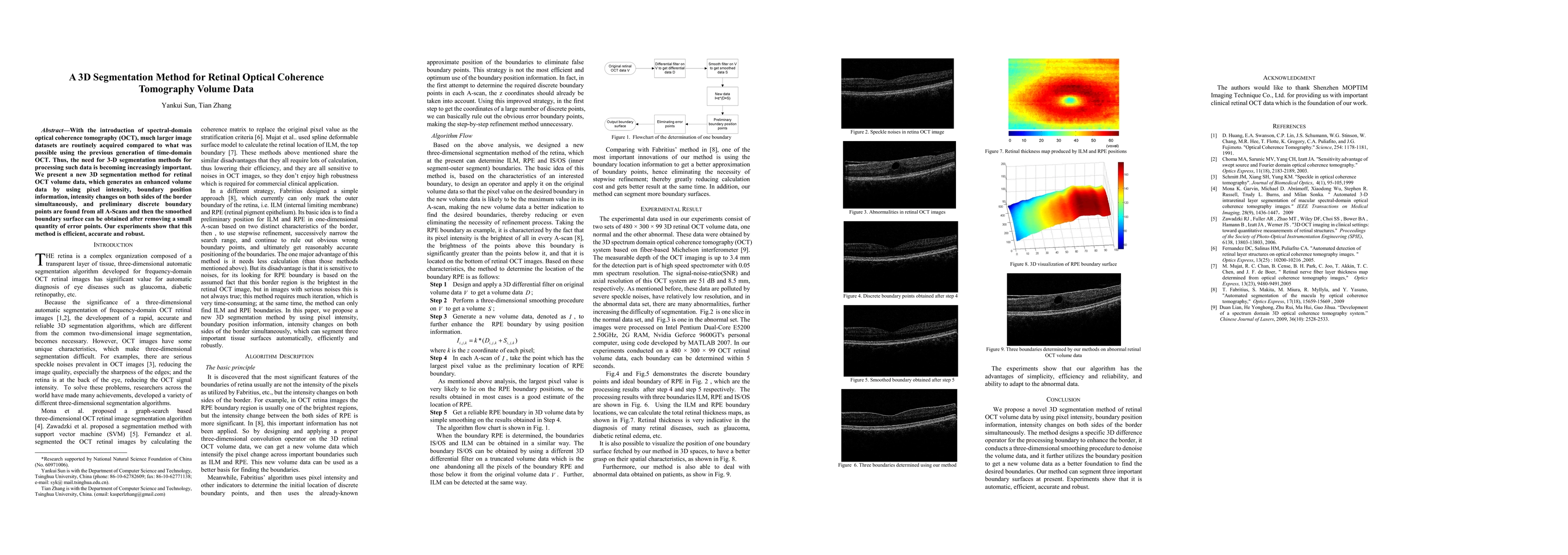
With the introduction of spectral-domain optical coherence tomography (OCT), much larger image datasets are routinely acquired compared to what was possible using the previous generation of time-domain OCT. Thus, the need for 3-D segmentation methods for processing such data is becoming increasingly important. We present a new 3D segmentation method for retinal OCT volume data, which generates an enhanced volume data by using pixel intensity, boundary position information, intensity changes on both sides of the border simultaneously, and preliminary discrete boundary points are found from all A-Scans and then the smoothed boundary surface can be obtained after removing a small quantity of error points. Our experiments show that this method is efficient, accurate and robust.
AI Key Findings
Get AI-generated insights about this paper's methodology, results, significance, and more — seven facets brought into focus.
Impact
Paper Details
PDF Preview
Key Terms
Citation Network
Current paper (gray), citations (green), references (blue)
Display is limited for performance on very large graphs.
Discussion 0