ArxivLens

Quick Summary:

This paper proposes a multi-decoder architecture for automated drusen segmentation in retinal OCT scans, treating it as a multitask learning problem. The method outperforms baselines by segmenting the OBRPE and BM layers separately and using an extra decoder for the area between them, with connections enhancing regularization.

arXiv Id: 1906.07679
Comments: Accepted for publication in MICCAI 2019
Date Published: 2019-07-25
Date Updated: 2019-07-25

Categories:

Key Terms (TF-IDF):

segmentation (0.302)
decoder (0.299)
respectively (0.166)
task (0.163)
membrane (0.161)
surrogate (0.159)
volumes (0.157)
class (0.150)
tomography (0.145)
validated (0.144)
aiming (0.144)
private (0.141)
evaluations (0.137)
data sets (0.136)
baselines (0.136)
interfaces (0.135)
outer (0.133)
automated (0.133)
coherence (0.132)
regularization (0.131)
License: http://arxiv.org/licenses/nonexclusive-distrib/1.0/

Summary:

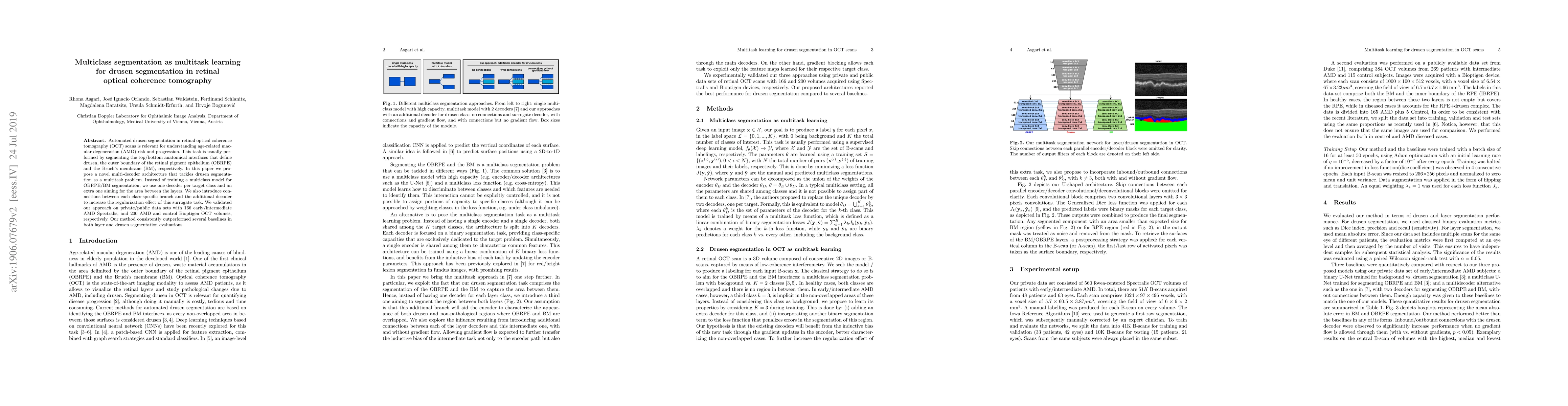
Automated drusen segmentation in retinal optical coherence tomography (OCT) scans is relevant for understanding age-related macular degeneration (AMD) risk and progression. This task is usually performed by segmenting the top/bottom anatomical interfaces that define drusen, the outer boundary of the retinal pigment epithelium (OBRPE) and the Bruch's membrane (BM), respectively. In this paper we propose a novel multi-decoder architecture that tackles drusen segmentation as a multitask problem. Instead of training a multiclass model for OBRPE/BM segmentation, we use one decoder per target class and an extra one aiming for the area between the layers. We also introduce connections between each class-specific branch and the additional decoder to increase the regularization effect of this surrogate task. We validated our approach on private/public data sets with 166 early/intermediate AMD Spectralis, and 200 AMD and control Bioptigen OCT volumes, respectively. Our method consistently outperformed several baselines in both layer and drusen segmentation evaluations.

Citation & Reference Network Visualization

This graph shows how this paper is connected to other research. The current paper is shown in dark gray, papers that cite it are in green, and papers it references are in blue. You can:

Current Paper
Papers citing this work (Citations)
Papers cited by this work (References)

• Hover over any node to see the paper title

• Click a node to view that paper's details

• Drag nodes to rearrange the graph

• Use the zoom controls (bottom right) to zoom in/out

Citations and References

Title Authors Year Actions
Targeted Microperimetry Grids for Focal Lesions in Intermediate AMD: PINNACLE Study Report 7
Stefan Futterknecht, Philipp Anders, Julia Mai, Sophie Riedl, Ursula Hall, Chrysoula Gabrani, Kristina Pfau, M. Menten, Daniel Rueckert, A. T. Prevost, H. Bogunović, Lars G Fritsche, U. Schmidt-Erfurth, S. Sivaprasad, A. Lotery, Hendrik P. N. Scholl, Maximilian Pfau
2025
View Paper
Multiscale attention-over-attention network for retinal disease recognition in OCT radiology images
Abdulmajeed M. Alenezi, Daniyah A. Aloqalaa, Sushil Kumar Singh, Raqinah Alrabiah, Shabana Habib, Muhammad Islam, Y. Daradkeh
2024
View Paper
Sequence of Morphological Changes Preceding Atrophy in Intermediate AMD Using Deep Learning
Sophie Riedl, U. Schmidt-Erfurth, Antoine Rivail, Klaudia Birner, Julia Mai, Wolf-Dieter Vogl, Zhichao Wu, Robyn H Guymer, H. Bogunović, G. Reiter
2024
View Paper
Identifying Ocular Diseases through Image Processing: Pattern Matching on Single Fundus Images
Simona Correra, Giulia Varriano, Vittoria Nardone, A. Santone
2024
View Paper
ADVANCES IN PHOTORECEPTOR AND RETINAL PIGMENT EPITHELIUM QUANTIFICATIONS IN INTERMEDIATE AGE-RELATED MACULAR DEGENERATION
Sophie Frank, G. Reiter, Oliver Leingang, Philipp Fuchs, L. Coulibaly, Virginia Mares, H. Bogunović, U. Schmidt-Erfurth
2024
View Paper
Classification and segmentation of OCT images for age-related macular degeneration based on dual guidance networks
Shengyong Diao, Jinzhu Su, Changqing Yang, Weifang Zhu, Dehui Xiang, Xinjian Chen, Qing Peng, Fei Shi
2023
View Paper
Accurate drusen segmentation in optical coherence tomography via order-constrained regression of retinal layer heights
Olivier Morelle, M. Wintergerst, R. Finger, T. Schultz
2023
View Paper
Contrastive uncertainty based biomarkers detection in retinal optical coherence tomography images
Xiaoming Liu, Kejie Zhou, Junping Yao, Man Wang, Ying Zhang
2022
View Paper
Multi-Task Multi-Scale Learning For Outcome Prediction in 3D PET Images
Amine Amyar, R. Modzelewski, P. Vera, V. Morard, S. Ruan
2022
View Paper
Trustworthy AI: Closing the gap between development and integration of AI systems in ophthalmic practice
C. González-Gonzalo, E. Thee, C. Klaver, Aaron Y. Lee, R. Schlingemann, A. Tufail, F. Verbraak, C. I. Sánchez
2021
View Paper
MsTGANet: Automatic Drusen Segmentation From Retinal OCT Images
Meng Wang, Weifang Zhu, Fei Shi, Jinzhu Su, Haoyu Chen, Kai Yu, Yi Zhou, Yuanyuan Peng, Zhongyue Chen, Xinjian Chen
2021
View Paper
Weakly supervised anomaly segmentation in retinal OCT images using an adversarial learning approach.
Jing Wang, Wanyue Li, Yiwei Chen, Wangyi Fang, Wen Kong, Yi He, Guohua Shi
2021
View Paper
Spatio-temporal alterations in retinal and choroidal layers in the progression of age-related macular degeneration (AMD) in optical coherence tomography
Wolf-Dieter Vogl, H. Bogunović, S. Waldstein, Sophie Riedl, U. Schmidt-Erfurth
2021
View Paper
A Benchmark of Ocular Disease Intelligent Recognition: One Shot for Multi-disease Detection
Ning Li, Tao Li, Chunyu Hu, Kai Wang, Hong Kang
2021
View Paper
Topographic Distribution and Progression of Soft Drusen Volume in Age-Related Macular Degeneration Implicate Neurobiology of Fovea
A. Pollreisz, G. Reiter, H. Bogunović, L. Baumann, Astrid Jakob, F. Schlanitz, S. Sacu, C. Owsley, K. Sloan, C. Curcio, U. Schmidt-Erfurth
2021
View Paper
Automatic Drusen Segmentation for Age-Related Macular Degeneration in Fundus Images Using Deep Learning
Quang T. M. Pham, Sang-il Ahn, S. Song, Jitae Shin
2020
View Paper
MMCL-Net: Spinal disease diagnosis in global mode using progressive multi-task joint learning
Yanfei Hong, B. Wei, Zhongyi Han, Xiang Li, Yuanjie Zheng, Shuo Li
2020
View Paper
Unified generative adversarial networks for multimodal segmentation from unpaired 3D medical images
Wenguang Yuan, Jia Wei, Jiabing Wang, Qianli Ma, T. Tasdizen
2020
View Paper
Two-Stage Cross-Domain Ocular Disease Recognition With Data Augmentation
Qiong Wang, Zhilin Guo, Jun Yao, Nan Yan
2023
View Paper
Embedded Residual Recurrent Network and Graph Search for the Segmentation of Retinal Layer Boundaries in Optical Coherence Tomography
Kai Hu, Dong Liu, Zhineng Chen, Xuanya Li, Yuan Zhang, Xieping Gao
2021
View Paper
Deep learning for ophthalmology using optical coherence tomography
Henry A. Leopold, Amitojdeep Singh, S. Sengupta, V. Lakshminarayanan
2021
View Paper
CAMEL: Confidence-Aware Multi-task Ensemble Learning with Spatial Information for Retina OCT Image Classification and Segmentation
Juho Jung, Migyeong Yang, Hyunseon Won, Jiwon Kim, Jeong Mo, J. Hwang, Daniel Duck-Jin, Duck-Jin Hwang, Jinyoung Han
View Paper

Comments (0)

This website uses cookies to ensure you get the best experience. Learn more