This paper proposes an enhanced U-Net with spatial pyramid pooling for segmenting drusen in optical coherence tomography (OCT) images, which outperforms the standard U-Net model. The network also segments the outer boundary retinal pigment epithelium and Bruch's membrane, aiding in the automated management of age-related macular degeneration.
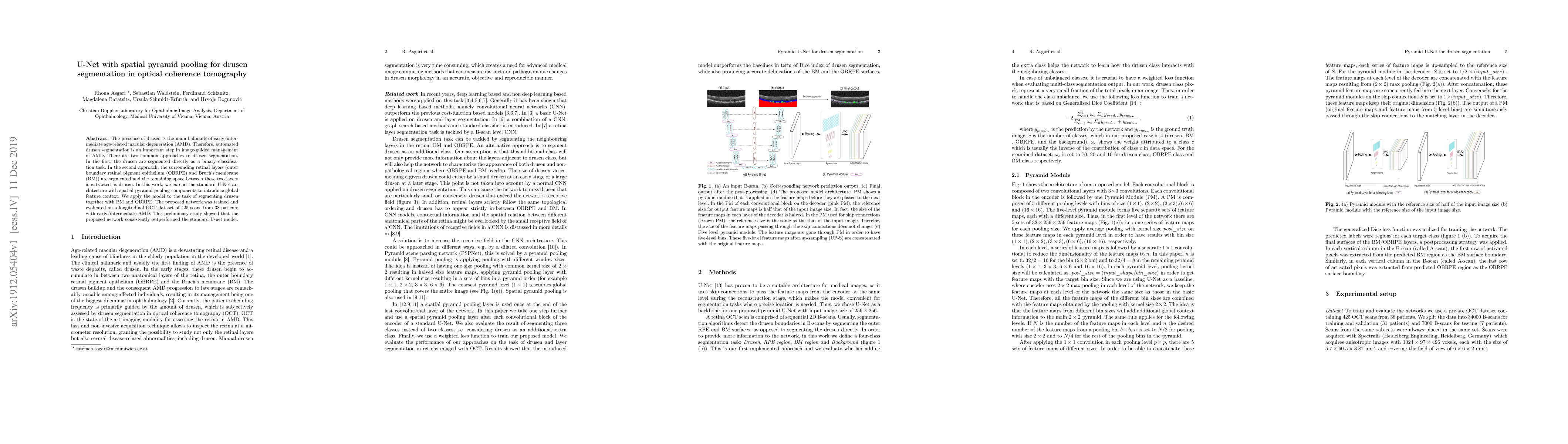
The presence of drusen is the main hallmark of early/intermediate age-related macular degeneration (AMD). Therefore, automated drusen segmentation is an important step in image-guided management of AMD. There are two common approaches to drusen segmentation. In the first, the drusen are segmented directly as a binary classification task. In the second approach, the surrounding retinal layers (outer boundary retinal pigment epithelium (OBRPE) and Bruch's membrane (BM)) are segmented and the remaining space between these two layers is extracted as drusen. In this work, we extend the standard U-Net architecture with spatial pyramid pooling components to introduce global feature context. We apply the model to the task of segmenting drusen together with BM and OBRPE. The proposed network was trained and evaluated on a longitudinal OCT dataset of 425 scans from 38 patients with early/intermediate AMD. This preliminary study showed that the proposed network consistently outperformed the standard U-net model.
This graph shows how this paper is connected to other research. The current paper is shown in dark gray, papers that cite it are in green, and papers it references are in blue. You can:
• Hover over any node to see the paper title
• Click a node to view that paper's details
• Drag nodes to rearrange the graph
• Use the zoom controls (bottom right) to zoom in/out
Title | Authors | Year | Actions |
---|---|---|---|
Technology for detection and subtype classification of drusen using OCT data for diagnosing age-related macular degeneration
|
N. Ilyasova, N. Demin, D.V. Kirsh
|
2024
|
View Paper |
Lesion Detection in Optical Coherence Tomography with Transformer-Enhanced Detector
|
H. Ahmed, Qianni Zhang, Ferranti Wong, Robert Donnan, Akram Alomainy
|
2023
|
View Paper |
Deep Learning with Automatic Data Augmentation for Segmenting Schisis Cavities in the Optical Coherence Tomography Images of X-Linked Juvenile Retinoschisis Patients
|
Xing Wei, Hui Li, T. Zhu, Wuyi Li, Yamei Li, R. Sui
|
2023
|
View Paper |
Accurate drusen segmentation in optical coherence tomography via order-constrained regression of retinal layer heights
|
O. Morelle, M. Wintergerst, R. Finger, T. Schultz
|
2023
|
View Paper |
Medical Image Segmentation Review: The Success of U-Net
|
Reza Azad, Ehsan Khodapanah Aghdam, Amelie Rauland, Yiwei Jia, Atlas Haddadi Avval, Afshin Bozorgpour, Sanaz Karimijafarbigloo, J. P. Cohen, E. Adeli, D. Merhof
|
2022
|
View Paper |
A Review on Multiscale-Deep-Learning Applications
|
Elizar Elizar, M. A. Zulkifley, R. Muharar, M. H. M. Zaman, S. M. Mustaza
|
2022
|
View Paper |
MsTGANet: Automatic Drusen Segmentation From Retinal OCT Images
|
Meng Wang, Weifang Zhu, Fei Shi, Jinzhu Su, Haoyu Chen, Kai Yu, Yi Zhou, Yuanyuan Peng, Zhongyue Chen, Xinjian Chen
|
2021
|
View Paper |
Automated segmentation of macular edema for the diagnosis of ocular disease using deep learning method
|
Zhenhua Wang, Yu-Ling Zhong, Mudi Yao, Yan Ma, Wenping Zhang, Chaopeng Li, Z. Tao, Qin Jiang, B. Yan
|
2021
|
View Paper |
A Refined Pyramid Scene Parsing Network for Polarimetric SAR Image Semantic Segmentation in Agricultural Areas
|
Rui Zhang, Jie Chen, Liang Feng, Shuang Li, Wei Yang, Ding Guo
|
2021
|
View Paper |
A measurement of epidermal thickness of fingertip skin from OCT images using convolutional neural network
|
Yongping Lin, Dezi Li, Wang Liu, Z. Zhong, Zhifang Li, Youwu He, Shulian Wu
|
2020
|
View Paper |
U-Net and Its Variants for Medical Image Segmentation: A Review of Theory and Applications
|
N. Siddique, Sidike Paheding, Colin P. Elkin, V. Devabhaktuni
|
2020
|
View Paper |
Survey on Convolutional Neural Network Pooling Methods
|
袁铭阳, 周长胜, 黄宏博, 胡志颖
|
2020
|
View Paper |
An unsupervised hierarchical approach for automatic intra-retinal cyst segmentation in spectral-domain optical coherence tomography images.
|
Razieh Ganjee, M. Ebrahimi Moghaddam, R. Nourinia
|
2020
|
View Paper |
ADR-Net: Context Extraction Network based on M-Net for Medical Image Segmentation.
|
L. Ji, Xiaoyan Jiang, Yongbin Gao, Zhijun Fang, Q. Cai, Ziran Wei
|
2020
|
View Paper |
Retinal OCT Analysis and Prediction with Deep Learning
|
H. Bogunović, Wolf-Dieter Vogl, Philipp Seeböck, Antoine Rivail, Úrsula
|
2020
|
View Paper |
Joint Segmentation and Quantification of Chorioretinal Biomarkers in Optical Coherence Tomography Scans: A Deep Learning Approach
|
Bilal Hassan, S. Qin, Taimur Hassan, Ramsha Ahmed, N. Werghi
|
2021
|
View Paper |
Deep learning for ophthalmology using optical coherence tomography
|
Henry A. Leopold, Amitojdeep Singh, S. Sengupta, V. Lakshminarayanan
|
2021
|
View Paper |
Impact of Data Augmentation on Retinal OCT Image Segmentation for Diabetic Macular Edema Analysis
|
Daniel Bar-David, Laura Bar-David, Shiri Soudry, A. Fischer
|
2021
|
View Paper |
Published: 2019-07-25
Published: 2021-02-09
Comments (0)