01
MethodologyHow they did it
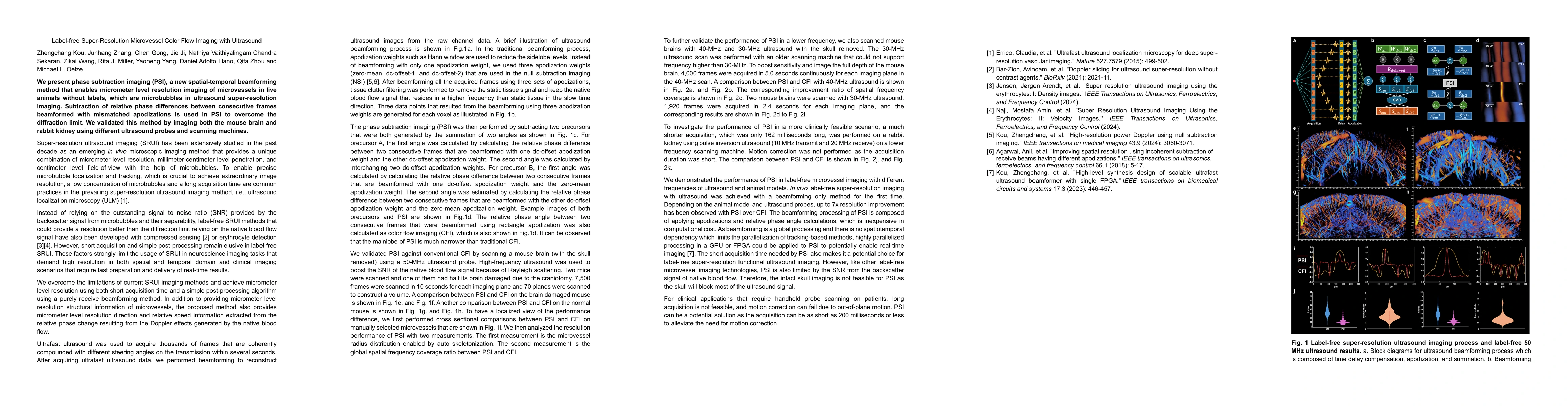
The research introduces phase subtraction imaging (PSI), a novel spatial-temporal beamforming method that achieves micrometer-level resolution imaging of microvessels in live animals without the use of labels, unlike conventional ultrasound super-resolution imaging that relies on microbubbles.
Discussion 0