Quick Summary:

This paper proposes a morphological reconstruction method to enhance microvessel mapping in super-resolution ultrasound imaging, achieving a fourfold increase in peak detection and a six-fold improvement in spatial resolution. This approach is computationally efficient and robust to noise, potentially advancing clinical translation of super-resolution ultrasound technology.

Summary

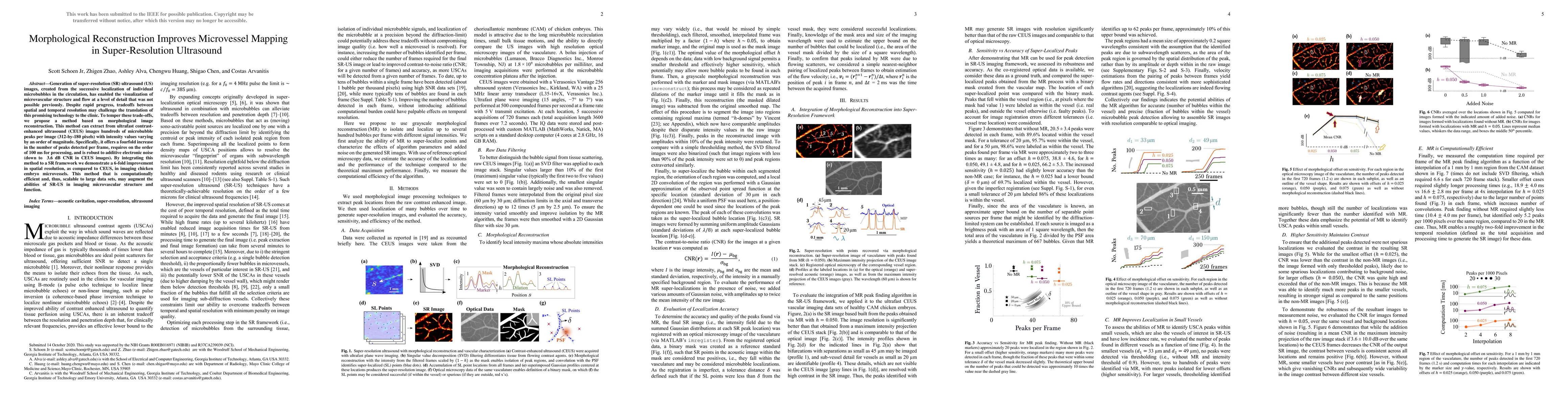
Generation of super-resolution (SR) ultrasound (US) images, created from the successive local-ization of individual microbubbles in the circulation, has enabled the visualization of microvascular structure and flow at a level of detail that was not possible previously. Despite rapid progress, tradeoffs between spatial and temporal resolution may challenge the translation of this promising technology to the clinic. To temper these trade-offs, we propose a method based on morphological image reconstriction. This method can extract from ultrafast contrast-enhanced ultrasound (CEUS) images hundreds of microbubble peaks per image (312-by-180 pixels) with intensity values varying by an order of magnitude. Specifically, it offers a fourfold increase in the number of peaks detected per frame, requires on the order of 100 ms for processing, and is robust to additive electronic noise (down to 3.6 dB CNR in CEUS images). By integrating this method to a SR framework we demonstrate a 6-fold improvement in spatial resolution, as compared to CEUS, in imaging chicken embryo microvessels. This method that is computationally efficient and, thus, scalable to large data sets, may augment the abilities of SR-US in imaging microvascular structure and function.

AI Key Findings

Get AI-generated insights about this paper's methodology, results, and significance.

Paper Details

PDF Preview

Download PDF

Key Terms

sr (0.285)
ultrasound (0.261)
peaks (0.201)
images (0.183)
resolution (0.180)
imaging (0.167)
spatial (0.148)
image (0.145)
method (0.139)
db (0.137)
morphological (0.136)
super resolution (0.135)
successive (0.135)
ms (0.132)
enabled (0.129)
additive (0.128)
computationally efficient (0.128)
trade (0.120)
data sets (0.120)
fold (0.120)
Title Authors Year Actions
Learning-based super-resolution of 3D ultrasound images
Simone Cammarasana, Giuseppe Patané
2025
View Paper
Super-Resolution Ultrasound: From Data Acquisition and Motion Correction to Localization, Tracking, and Evaluation
S. Dencks, Mathew R. Lowerison, Joseph Hansen-Shearer, YiRang Shin, Georg Schmitz, Peng Song, Meng-Xing Tang
2025
View Paper
Microbubble Contrast Agents Improve Detection of Active Hemorrhage
Scott Schoen Jr, Alexis Prasov, Ion Candel, Saaid H. Arshad, M. Ottensmeyer, Theodore T. Pierce, L. Brattain, Brian A. Telfer, Anthony E. Samir
2024
View Paper
ULTRA-SR Challenge: Assessment of Ultrasound Localization and TRacking Algorithms for Super-Resolution Imaging
M. Lerendegui, K. Riemer, Georgios K. Papageorgiou, Bingxue Wang, Lachlan Arthur, Arthur Chavignon, Tao Zhang, Olivier Couture, Pingtong Huang, Md Ashikuzzaman, S. Dencks, C. Dunsby, Brandon Helfield, Jørgen Arendt Jensen, Thomas Lisson, Matthew R. Lowerison, H. Rivaz, Anthony E. Samir, Georg Schmitz, Scott Schoen, Ruud J. G. van Sloun, Peng Song, Tristan S. W. Stevens, Jipeng Yan, V. Sboros, Meng-Xing Tang
2024
View Paper
Study of a graphene optical sensor applied in glottic tumor
Jacilene M. Medeiros, F. C. Rufino, A. Montagnoli, G. Cirino, J. A. Diniz
2023
View Paper
Super-resolution of 2D ultrasound images and videos
Simone Cammarasana, Paolo Nicolardi, G. Patané
2023
View Paper
Assessment of Transarterial Chemoembolization Using Super-resolution Ultrasound Imaging and a Rat Model of Hepatocellular Carcinoma.
Katherine G. Brown, Jianing Li, Ryan Margolis, Brian G. Trinh, J. Eisenbrey, K. Hoyt
2023
View Paper
Robust Super-Resolution Ultrasound Microbubble Tracking with Optical Flow Guided Kalman Filter
Su-Lan Pu, Haolun Guo, Hui-Wen Xie, Guangquan Zhou
2022
View Paper
MR for ULTRA-SR: Improved Localization with Morphological Image Processing
S. Schoen, A. Samir, Viksit Kumar
2022
View Paper

Comments (0)

This website uses cookies to ensure you get the best experience. Learn more