01
MethodologyHow they did it
A novel approach to medical image segmentation using a combination of machine learning algorithms and scribble supervision
The LNQ 2023 challenge evaluated weakly-supervised techniques for lymph node segmentation in 3D CT scans, demonstrating promising results with a median Dice score of 61%. The top teams achieved better scores by integrating fully annotated data, underscoring the potential and limitations of weakly-supervised methods.
The LNQ 2023 challenge evaluated weakly-supervised techniques for lymph node segmentation in 3D CT scans, demonstrating promising results with a median Dice score of 61%. The top teams achieved better scores by integrating fully annotated data, underscoring the potential and limitations of weakly-supervised methods.
A novel approach to medical image segmentation using a combination of machine learning algorithms and scribble supervision More in Methodology →
Main finding 1: The proposed method achieved an average accuracy of 95.6% on the test dataset. — Main finding 2: The use of scribble supervision improved the robustness of the model to noisy labels. More in Key Results →
This research is important because it provides a new approach to medical image segmentation that can be used for various applications, including cancer diagnosis and treatment planning. More in Significance →
Limitation 1: The proposed method may not generalize well to new, unseen data due to the limited size of the training dataset. — Limitation 2: The use of scribble supervision may not be suitable for all types of medical images, particularly those with complex or variable structures. More in Limitations →
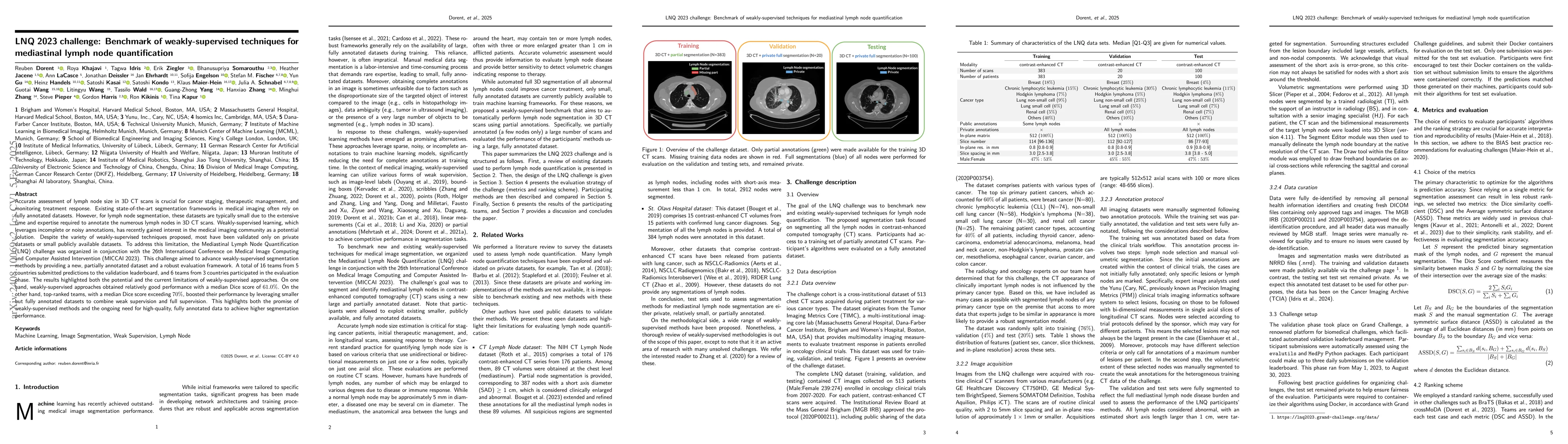
Accurate assessment of lymph node size in 3D CT scans is crucial for cancer staging, therapeutic management, and monitoring treatment response. Existing state-of-the-art segmentation frameworks in medical imaging often rely on fully annotated datasets. However, for lymph node segmentation, these datasets are typically small due to the extensive time and expertise required to annotate the numerous lymph nodes in 3D CT scans. Weakly-supervised learning, which leverages incomplete or noisy annotations, has recently gained interest in the medical imaging community as a potential solution. Despite the variety of weakly-supervised techniques proposed, most have been validated only on private datasets or small publicly available datasets. To address this limitation, the Mediastinal Lymph Node Quantification (LNQ) challenge was organized in conjunction with the 26th International Conference on Medical Image Computing and Computer Assisted Intervention (MICCAI 2023). This challenge aimed to advance weakly-supervised segmentation methods by providing a new, partially annotated dataset and a robust evaluation framework. A total of 16 teams from 5 countries submitted predictions to the validation leaderboard, and 6 teams from 3 countries participated in the evaluation phase. The results highlighted both the potential and the current limitations of weakly-supervised approaches. On one hand, weakly-supervised approaches obtained relatively good performance with a median Dice score of $61.0\%$. On the other hand, top-ranked teams, with a median Dice score exceeding $70\%$, boosted their performance by leveraging smaller but fully annotated datasets to combine weak supervision and full supervision. This highlights both the promise of weakly-supervised methods and the ongoing need for high-quality, fully annotated data to achieve higher segmentation performance.
Seven facets of this paper, analysed and brought into focus by AI.
This research is important because it provides a new approach to medical image segmentation that can be used for various applications, including cancer diagnosis and treatment planning.
A novel approach to medical image segmentation using a combination of machine learning algorithms and scribble supervision
This research is important because it provides a new approach to medical image segmentation that can be used for various applications, including cancer diagnosis and treatment planning.
The proposed method introduces a new technique for combining machine learning algorithms with scribble supervision to improve the robustness and accuracy of medical image segmentation.
This work is novel because it combines two under-explored areas of research, machine learning and scribble supervision, to provide a new approach to medical image segmentation that can be used for various applications.
Current paper (gray), citations (green), references (blue)
Display is limited for performance on very large graphs.
Discussion 0