Academic Profile
Statistics
Similar Authors
Papers on arXiv
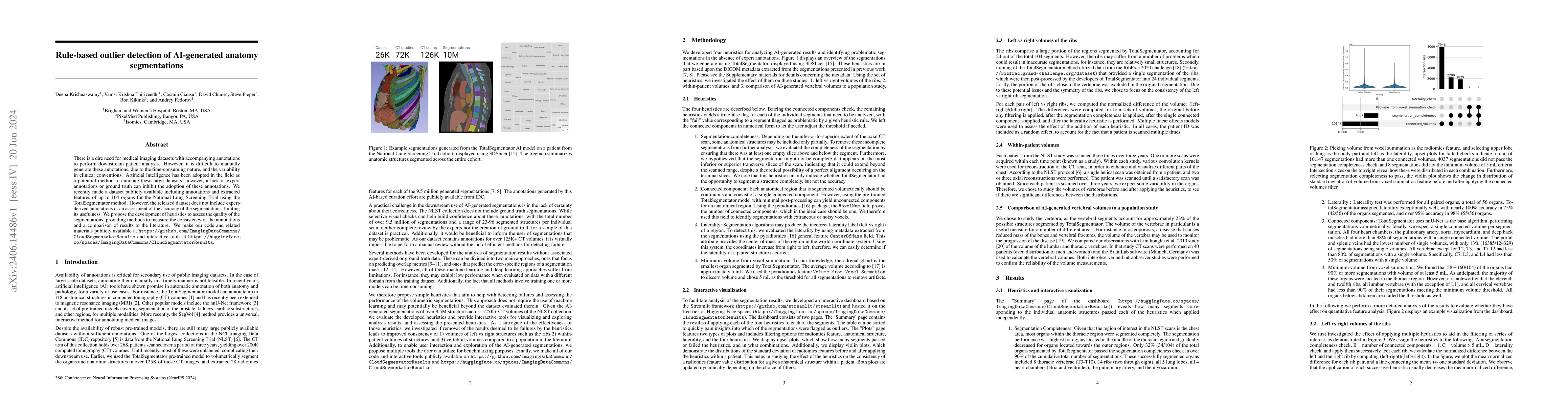
There is a dire need for medical imaging datasets with accompanying annotations to perform downstream patient analysis. However, it is difficult to manually generate these annotations, due to the ti...
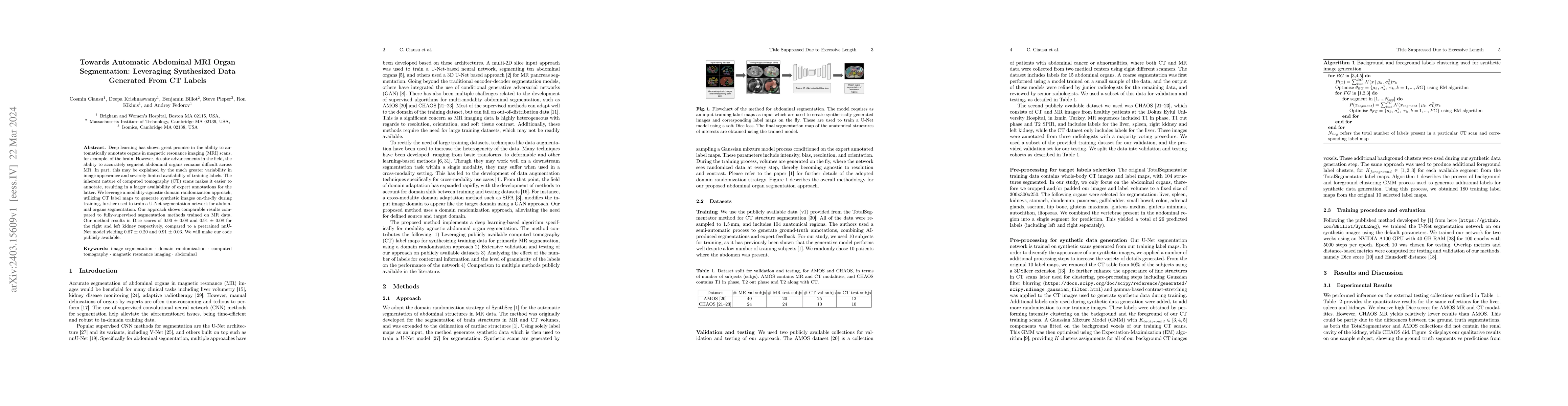
Deep learning has shown great promise in the ability to automatically annotate organs in magnetic resonance imaging (MRI) scans, for example, of the brain. However, despite advancements in the field...
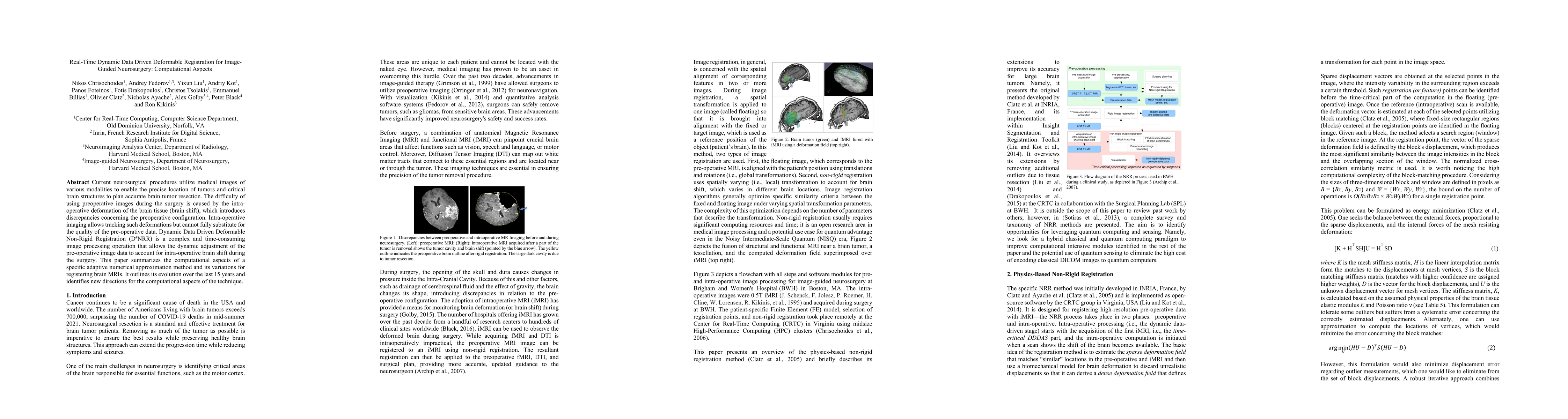
Current neurosurgical procedures utilize medical images of various modalities to enable the precise location of tumors and critical brain structures to plan accurate brain tumor resection. The diffi...

During neurosurgery, medical images of the brain are used to locate tumors and critical structures, but brain tissue shifts make pre-operative images unreliable for accurate removal of tumors. Intra...
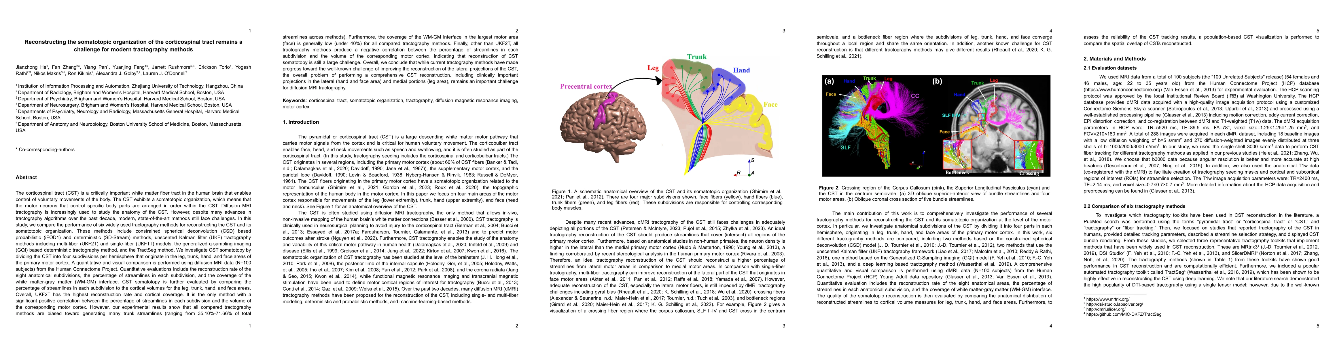
The corticospinal tract (CST) is a critically important white matter fiber tract in the human brain that enables control of voluntary movements of the body. Diffusion MRI tractography is the only me...
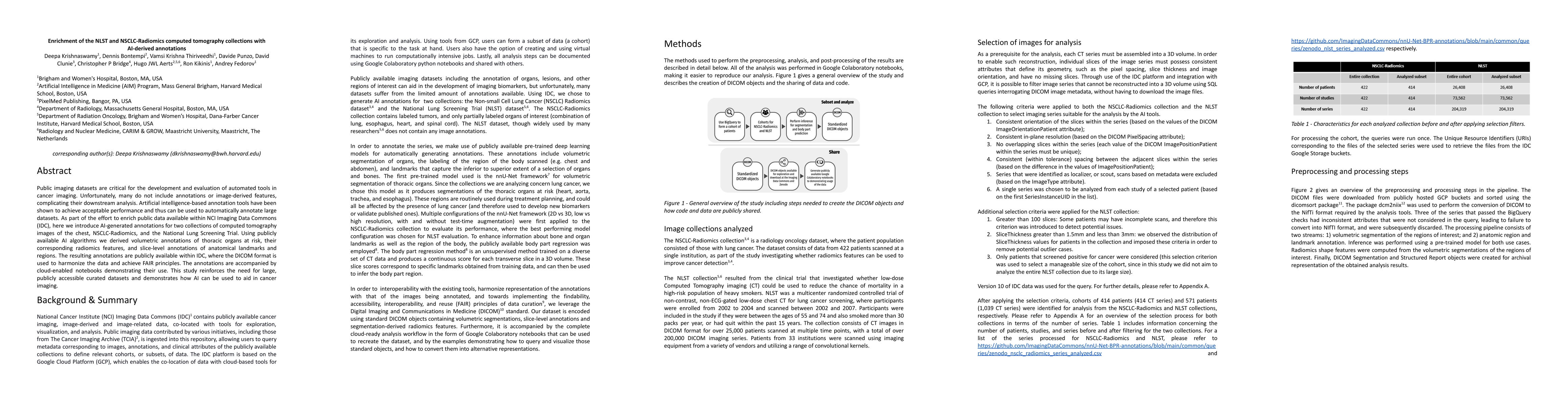
Public imaging datasets are critical for the development and evaluation of automated tools in cancer imaging. Unfortunately, many do not include annotations or image-derived features, complicating t...
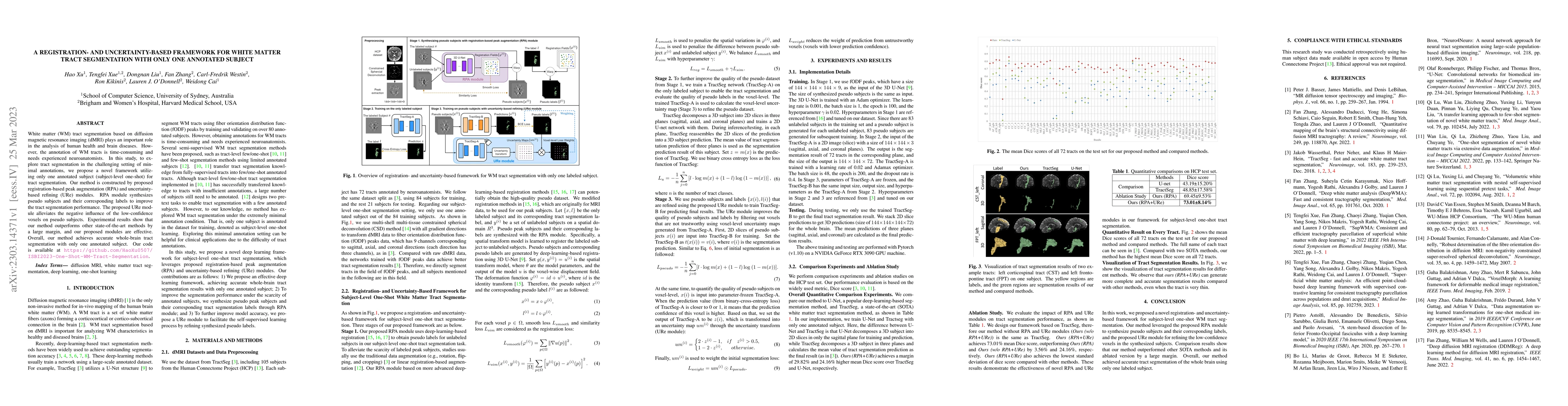
White matter (WM) tract segmentation based on diffusion magnetic resonance imaging (dMRI) plays an important role in the analysis of human health and brain diseases. However, the annotation of WM tr...
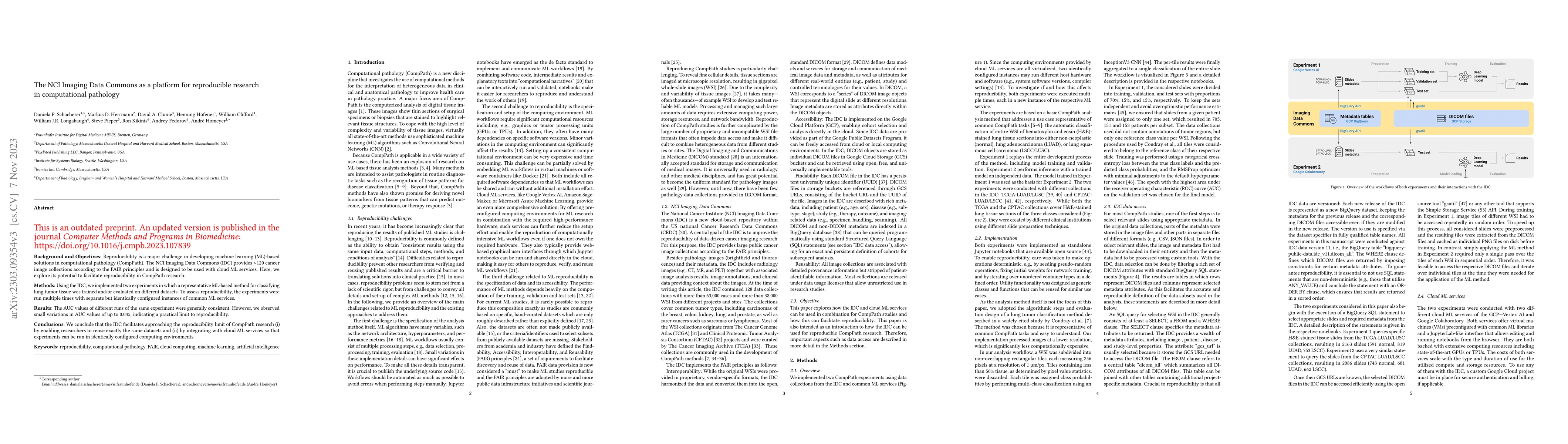
Background and Objectives: Reproducibility is a major challenge in developing machine learning (ML)-based solutions in computational pathology (CompPath). The NCI Imaging Data Commons (IDC) provides...
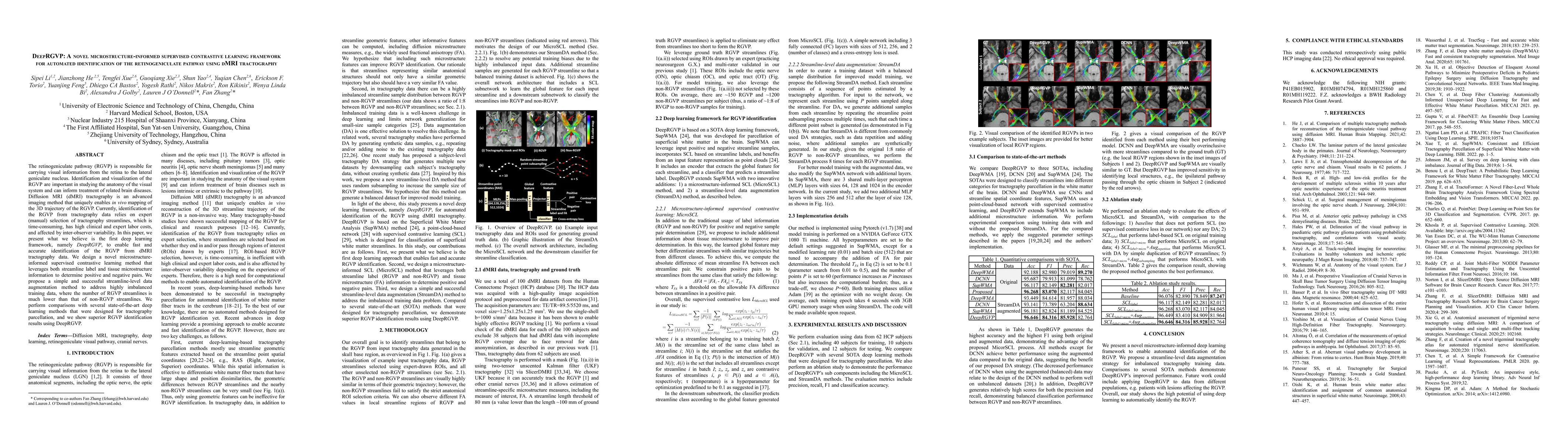
The retinogeniculate pathway (RGVP) is responsible for carrying visual information from the retina to the lateral geniculate nucleus. Identification and visualization of the RGVP are important in st...
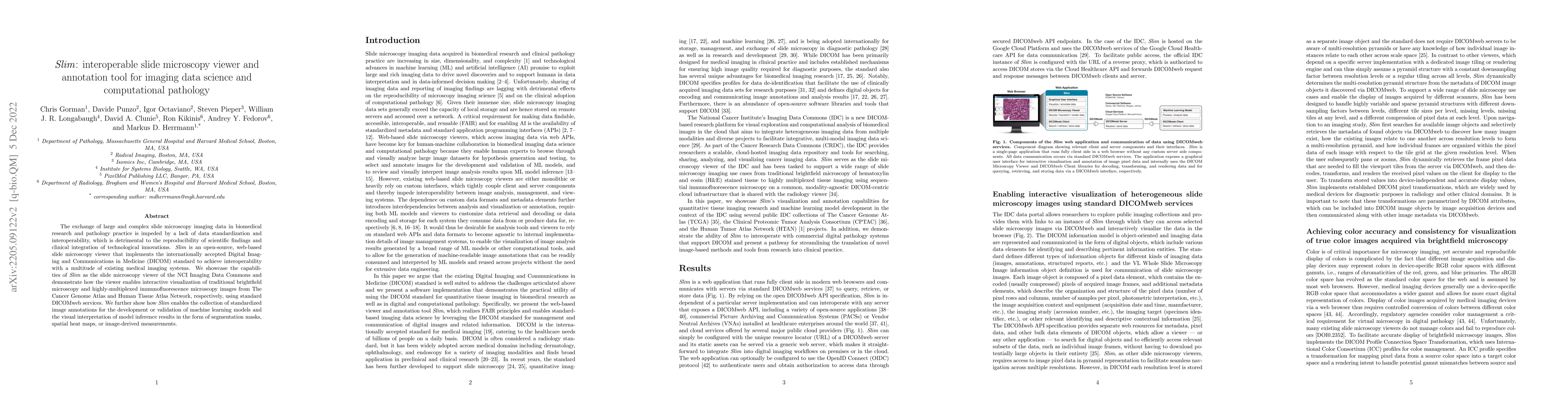
The exchange of large and complex slide microscopy imaging data in biomedical research and pathology practice is impeded by a lack of data standardization and interoperability, which is detrimental ...
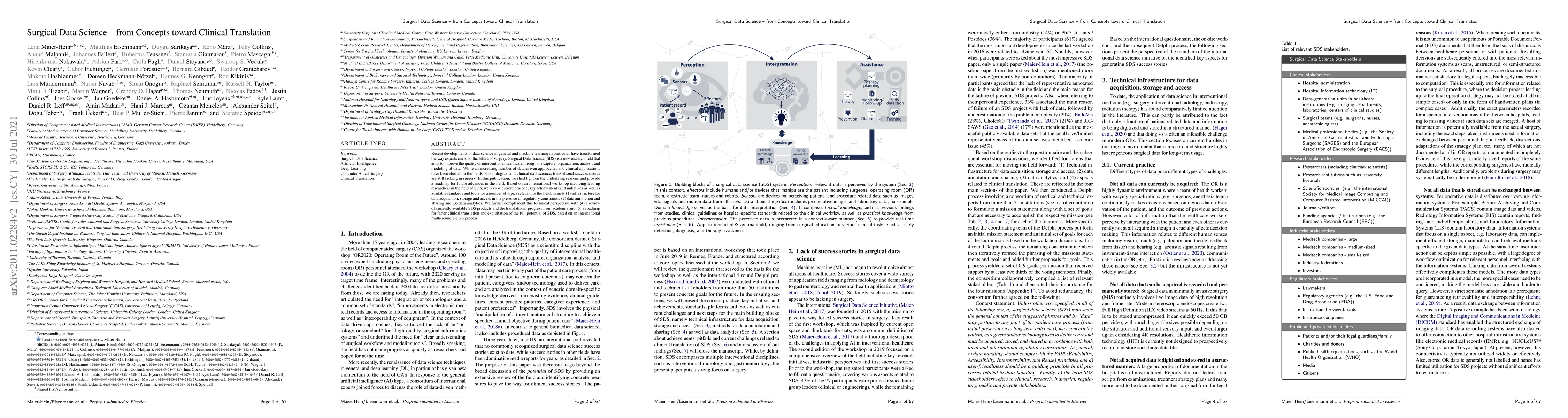
Recent developments in data science in general and machine learning in particular have transformed the way experts envision the future of surgery. Surgical Data Science (SDS) is a new research field...
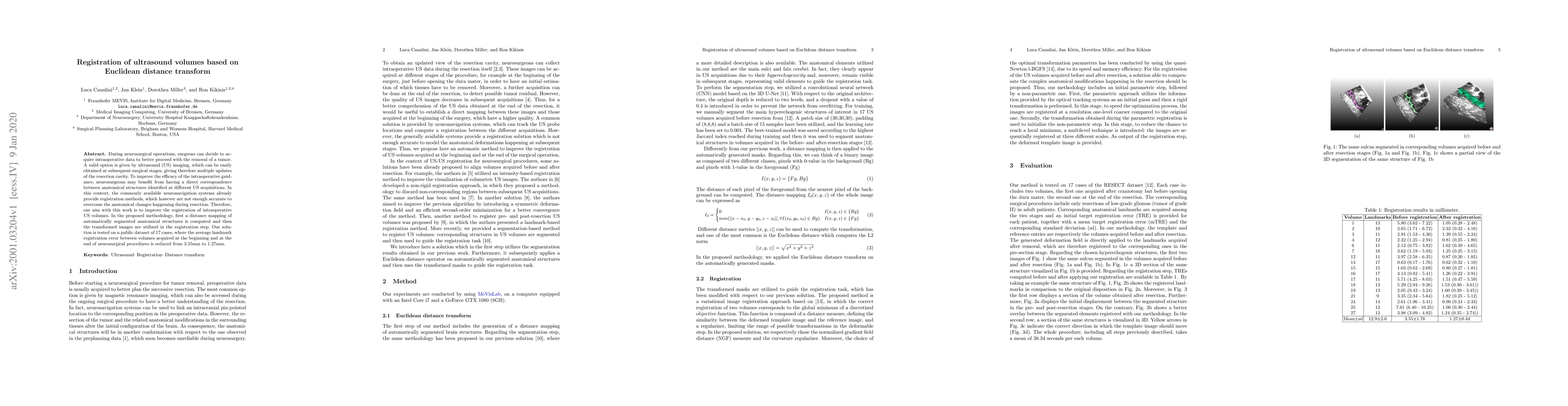
During neurosurgical operations, surgeons can decide to acquire intraoperative data to better proceed with the removal of a tumor. A valid option is given by ultrasound (US) imaging, which can be ea...
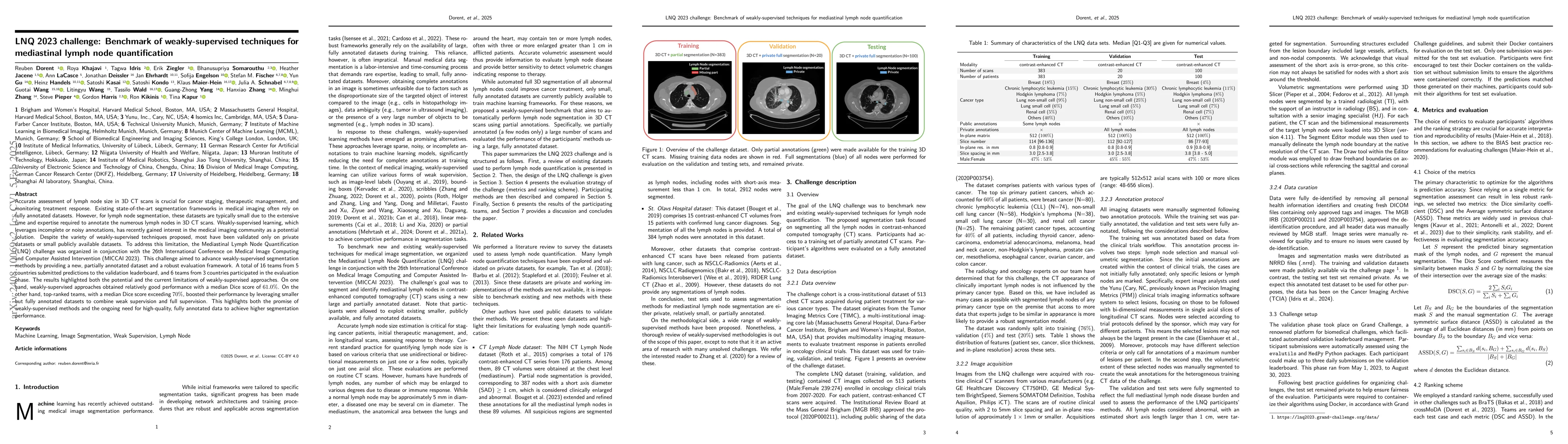
Accurate assessment of lymph node size in 3D CT scans is crucial for cancer staging, therapeutic management, and monitoring treatment response. Existing state-of-the-art segmentation frameworks in med...
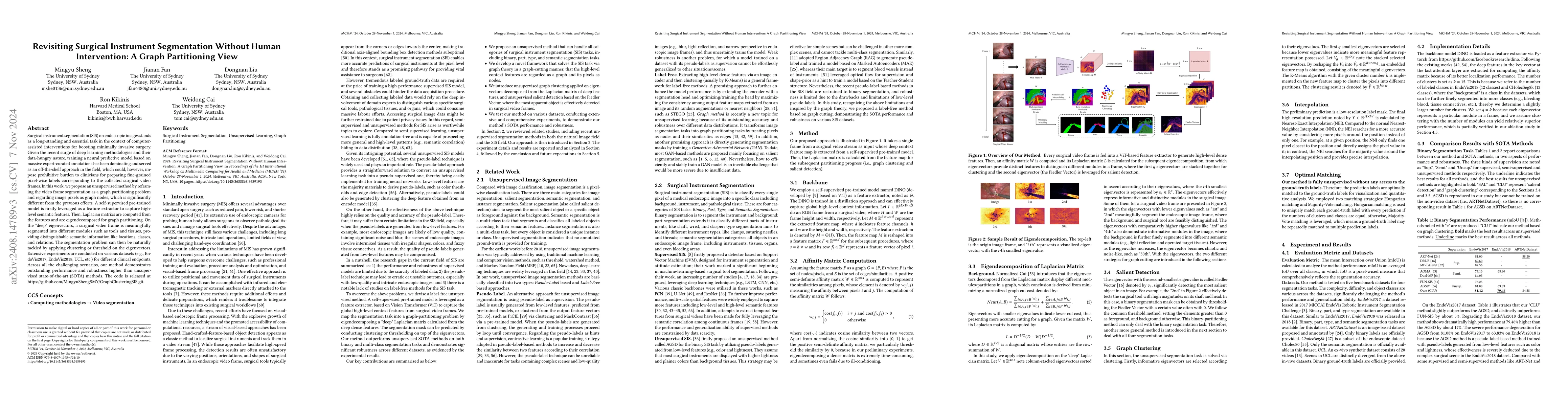
Surgical instrument segmentation (SIS) on endoscopic images stands as a long-standing and essential task in the context of computer-assisted interventions for boosting minimally invasive surgery. Give...
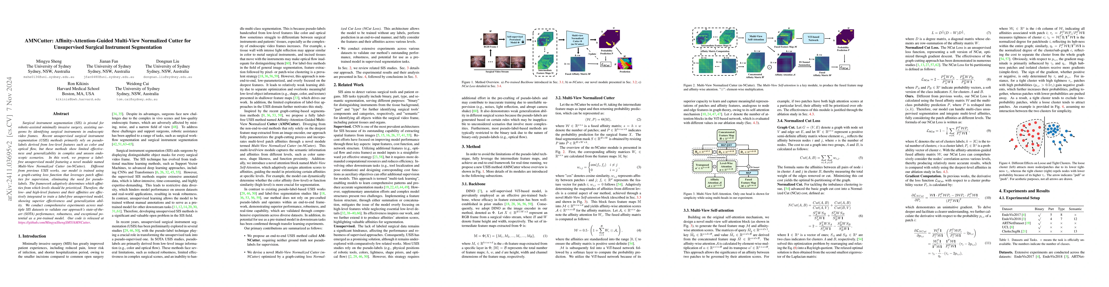
Surgical instrument segmentation (SIS) is pivotal for robotic-assisted minimally invasive surgery, assisting surgeons by identifying surgical instruments in endoscopic video frames. Recent unsupervise...
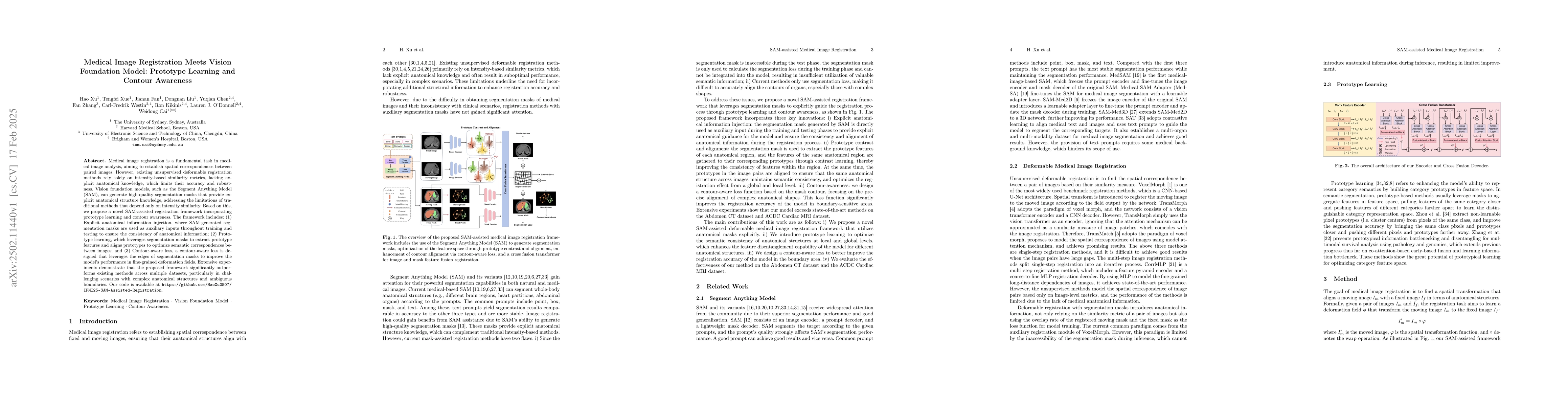
Medical image registration is a fundamental task in medical image analysis, aiming to establish spatial correspondences between paired images. However, existing unsupervised deformable registration me...
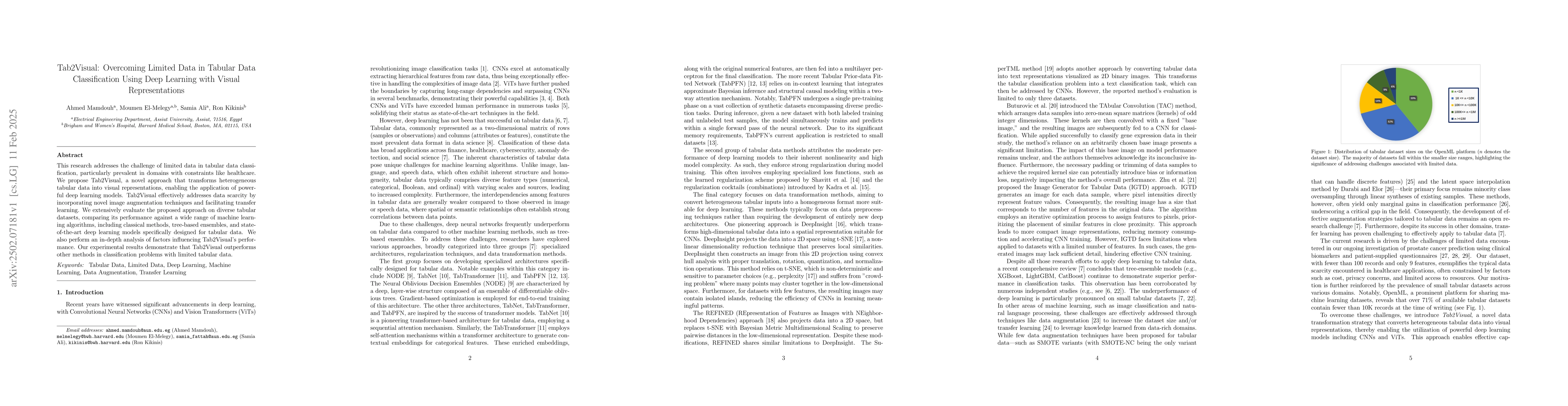
This research addresses the challenge of limited data in tabular data classification, particularly prevalent in domains with constraints like healthcare. We propose Tab2Visual, a novel approach that t...
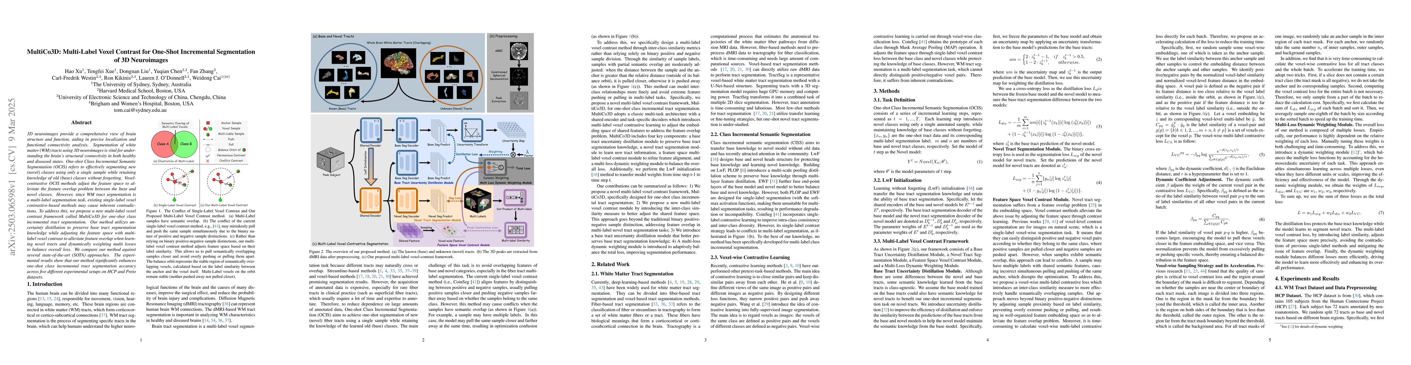
3D neuroimages provide a comprehensive view of brain structure and function, aiding in precise localization and functional connectivity analysis. Segmentation of white matter (WM) tracts using 3D neur...
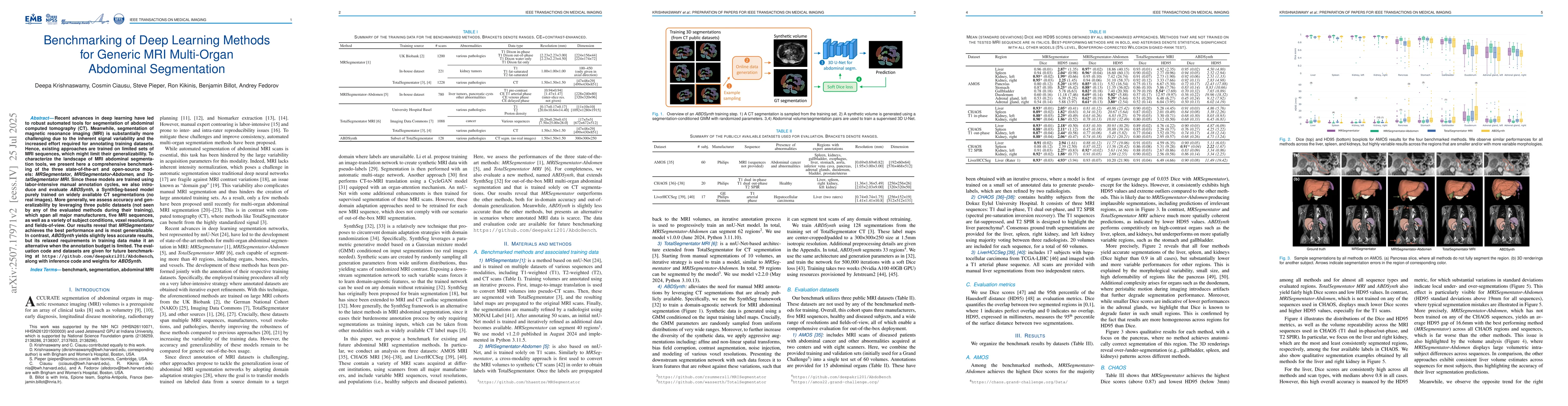
Recent advances in deep learning have led to robust automated tools for segmentation of abdominal computed tomography (CT). Meanwhile, segmentation of magnetic resonance imaging (MRI) is substantially...
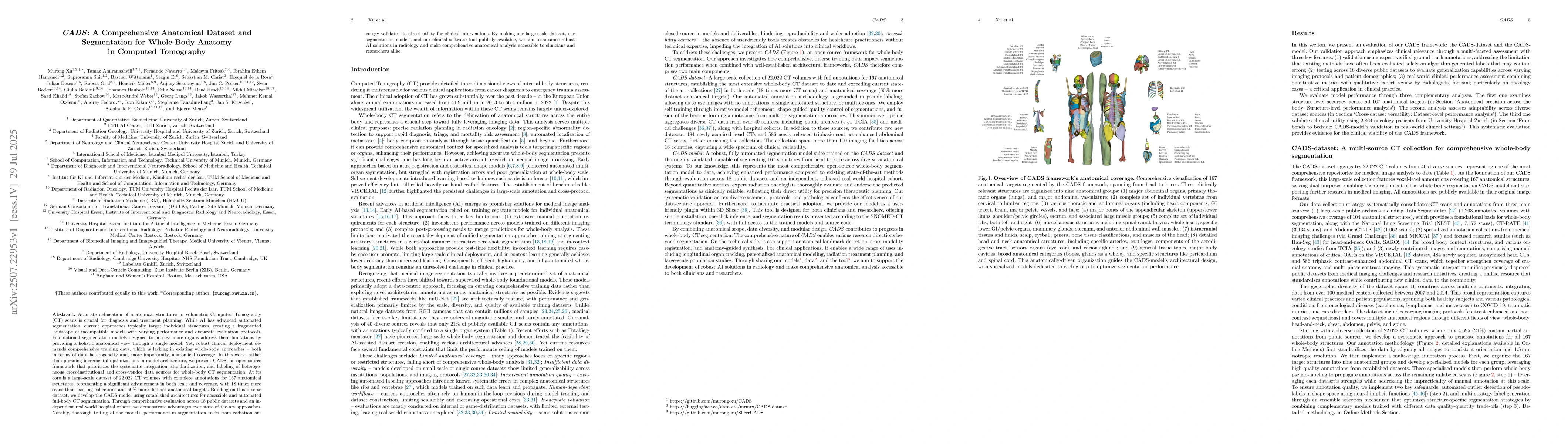
Accurate delineation of anatomical structures in volumetric CT scans is crucial for diagnosis and treatment planning. While AI has advanced automated segmentation, current approaches typically target ...
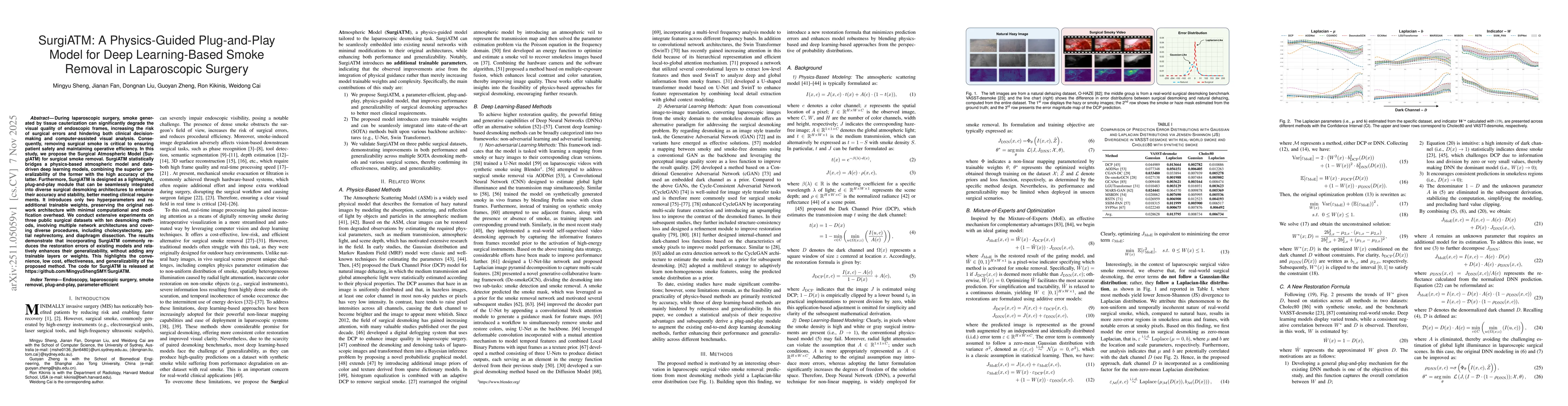
During laparoscopic surgery, smoke generated by tissue cauterization can significantly degrade the visual quality of endoscopic frames, increasing the risk of surgical errors and hindering both clinic...
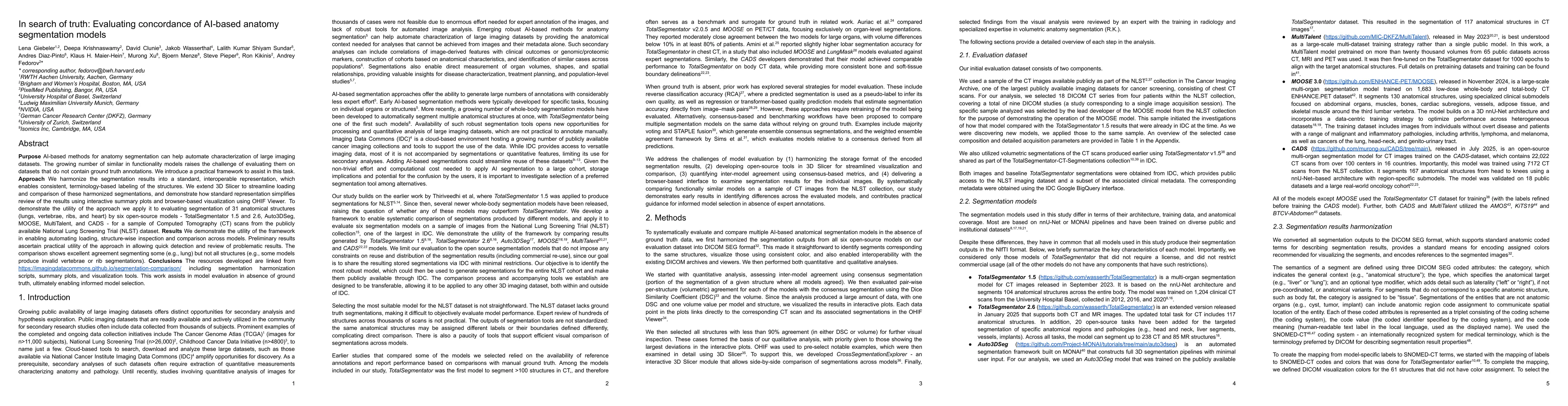
Purpose AI-based methods for anatomy segmentation can help automate characterization of large imaging datasets. The growing number of similar in functionality models raises the challenge of evaluating...
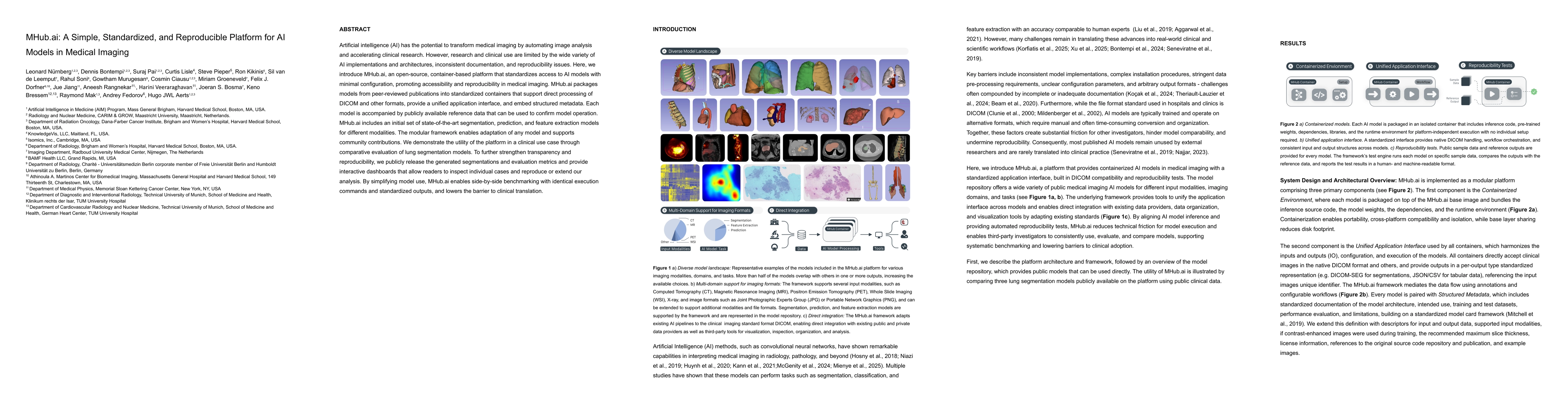
Artificial intelligence (AI) has the potential to transform medical imaging by automating image analysis and accelerating clinical research. However, research and clinical use are limited by the wide ...
In the last ten years, medical robotics has moved from the margins to the mainstream. Since the Engineering Research Center for Computer-Integrated Surgical Systems and Technology was Launched in 1998...