Revisiting Surgical Instrument Segmentation Without Human Intervention: A Graph Partitioning View
Publication
Metrics
AI Quick Summary
This research proposes an unsupervised method for surgical instrument segmentation in endoscopic images by framing it as a graph partitioning problem, eliminating the need for extensive human annotations. The method leverages self-supervised pre-trained models and Laplacian matrix eigendecomposition for segmentation, achieving superior performance and robustness compared to existing unsupervised techniques.
Paper Preview
Abstract
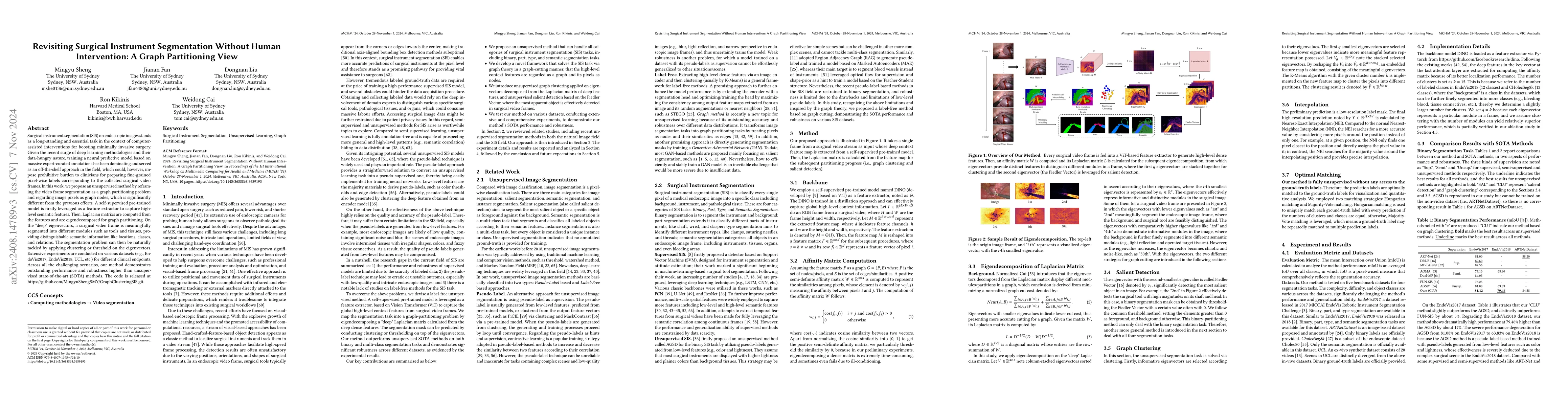
Surgical instrument segmentation (SIS) on endoscopic images stands as a long-standing and essential task in the context of computer-assisted interventions for boosting minimally invasive surgery. Given the recent surge of deep learning methodologies and their data-hungry nature, training a neural predictive model based on massive expert-curated annotations has been dominating and served as an off-the-shelf approach in the field, which could, however, impose prohibitive burden to clinicians for preparing fine-grained pixel-wise labels corresponding to the collected surgical video frames. In this work, we propose an unsupervised method by reframing the video frame segmentation as a graph partitioning problem and regarding image pixels as graph nodes, which is significantly different from the previous efforts. A self-supervised pre-trained model is firstly leveraged as a feature extractor to capture high-level semantic features. Then, Laplacian matrixs are computed from the features and are eigendecomposed for graph partitioning. On the "deep" eigenvectors, a surgical video frame is meaningfully segmented into different modules such as tools and tissues, providing distinguishable semantic information like locations, classes, and relations. The segmentation problem can then be naturally tackled by applying clustering or threshold on the eigenvectors. Extensive experiments are conducted on various datasets (e.g., EndoVis2017, EndoVis2018, UCL, etc.) for different clinical endpoints. Across all the challenging scenarios, our method demonstrates outstanding performance and robustness higher than unsupervised state-of-the-art (SOTA) methods. The code is released at https://github.com/MingyuShengSMY/GraphClusteringSIS.git.
AI Key Findings
Get AI-generated insights about this paper's methodology, results, significance, and more — seven facets brought into focus.
Impact
Authors
PDF Preview
Citation Network
Current paper (gray), citations (green), references (blue)
Display is limited for performance on very large graphs.
Discussion 0