Machine Learning for Analyzing Atomic Force Microscopy (AFM) Images Generated from Polymer Blends
Publication
Metrics
AI Quick Summary
This paper proposes a machine learning workflow using unsupervised techniques to automatically identify and segment polymer domains in AFM images, minimizing manual intervention. The discrete Fourier transform and cosine transform workflows performed better than ResNet50 deep learning for segmentation, and porespy was used to calculate domain size distributions, aiding in the characterization of polymer blends.
Paper Preview
Abstract
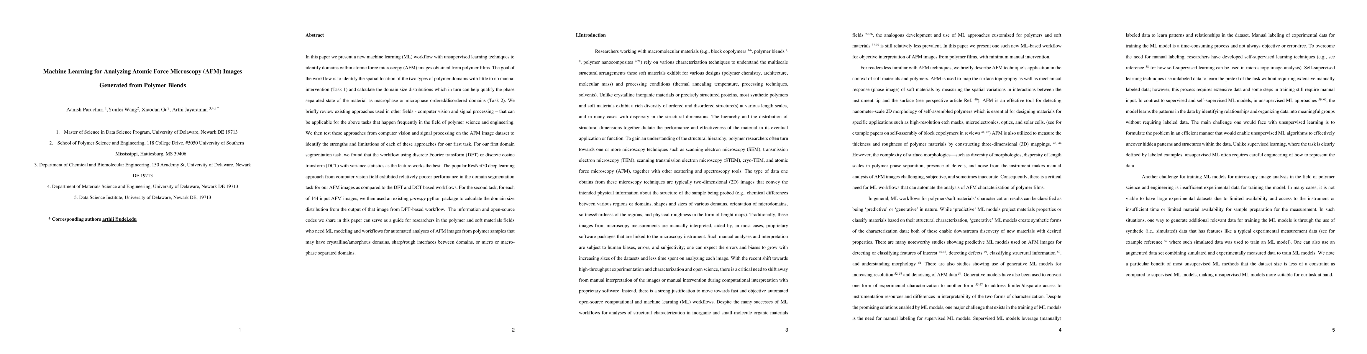
In this paper we present a new machine learning workflow with unsupervised learning techniques to identify domains within atomic force microscopy images obtained from polymer films. The goal of the workflow is to identify the spatial location of the two types of polymer domains with little to no manual intervention and calculate the domain size distributions which in turn can help qualify the phase separated state of the material as macrophase or microphase ordered or disordered domains. We briefly review existing approaches used in other fields, computer vision and signal processing that can be applicable for the above tasks that happen frequently in the field of polymer science and engineering. We then test these approaches from computer vision and signal processing on the AFM image dataset to identify the strengths and limitations of each of these approaches for our first task. For our first domain segmentation task, we found that the workflow using discrete Fourier transform or discrete cosine transform with variance statistics as the feature works the best. The popular ResNet50 deep learning approach from computer vision field exhibited relatively poorer performance in the domain segmentation task for our AFM images as compared to the DFT and DCT based workflows. For the second task, for each of 144 input AFM images, we then used an existing porespy python package to calculate the domain size distribution from the output of that image from DFT based workflow. The information and open source codes we share in this paper can serve as a guide for researchers in the polymer and soft materials fields who need ML modeling and workflows for automated analyses of AFM images from polymer samples that may have crystalline or amorphous domains, sharp or rough interfaces between domains, or micro or macrophase separated domains.
AI Key Findings
Get AI-generated insights about this paper's methodology, results, significance, and more — seven facets brought into focus.
Impact
Paper Details
Authors
PDF Preview
Citation Network
Current paper (gray), citations (green), references (blue)
Display is limited for performance on very large graphs.
Discussion 0