Methods to Quantify Dislocation Behavior with Dark-field X-ray Microscopy Timescans of Single-Crystal Aluminum
Publication
Metrics
AI Quick Summary
This paper introduces a semi-automated method to isolate, track, and quantify dislocation behavior using dark-field X-ray microscopy (DFXM) on single-crystal aluminum. The approach enables statistical characterization of dislocation shape, position, and motion, enhancing material models.
Paper Preview
Abstract
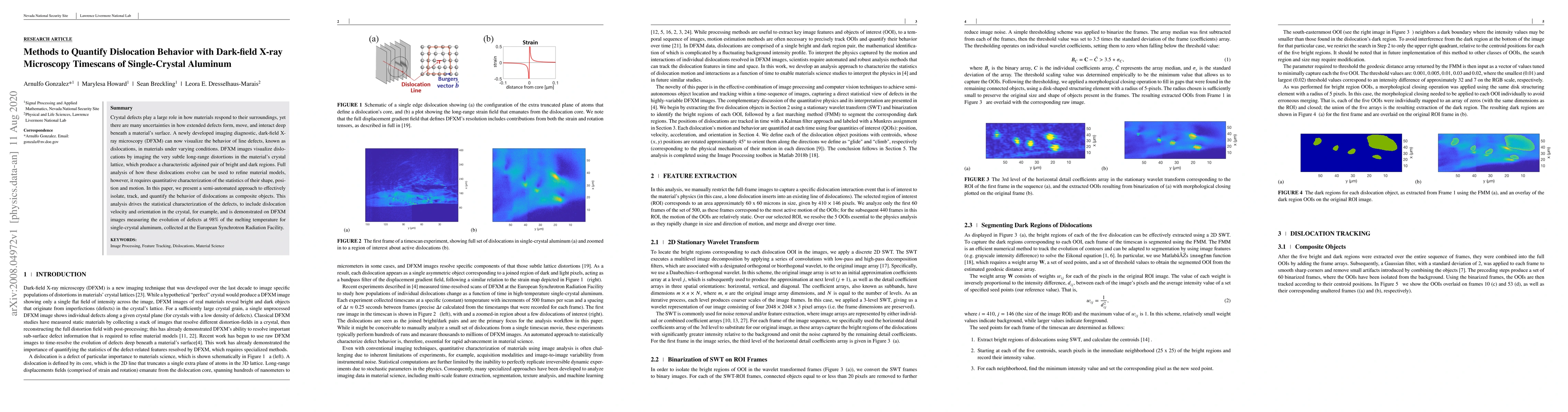
Crystal defects play a large role in how materials respond to their surroundings, yet there are many uncertainties in how extended defects form, move, and interact deep beneath a material's surface. A newly developed imaging diagnostic, dark-field X-ray microscopy (DFXM) can now visualize the behavior of line defects, known as dislocations, in materials under varying conditions. DFXM images visualize dislocations by imaging the very subtle long-range distortions in the material's crystal lattice, which produce a characteristic adjoined pair of bright and dark regions. Full analysis of how these dislocations evolve can be used to refine material models, however, it requires quantitative characterization of the statistics of their shape, position and motion. In this paper, we present a semi-automated approach to effectively isolate, track, and quantify the behavior of dislocations as composite objects. This analysis drives the statistical characterization of the defects, to include dislocation velocity and orientation in the crystal, for example, and is demonstrated on DFXM images measuring the evolution of defects at 98$\%$ of the melting temperature for single-crystal aluminum, collected at the European Synchrotron Radiation Facility.
AI Key Findings
Get AI-generated insights about this paper's methodology, results, significance, and more — seven facets brought into focus.
Impact
Paper Details
Authors
PDF Preview
Key Terms
Citation Network
Current paper (gray), citations (green), references (blue)
Display is limited for performance on very large graphs.
Discussion 0